Brachycephalic Upper Airway Obstruction Syndrome, BAOS, BAS, BUAOS
- Dr Andrew Matole, BVetMed, MSc

- Jun 18, 2022
- 6 min read

Brachycephalic Upper Airway Obstruction Syndrome (BUAOS) refers to an inherited respiratory disorder, a physical abnormality from birth that causes shortening of the bones of the skull when the soft tissues within the upper airways do not reduce in size proportionally plus the maldevelopment of the airway cartilages.
What are the clinical signs of Brachycephalic Upper Airway Obstruction Syndrome (BUAOS)?
The main clinical signs of Brachycephalic Upper Airway Obstruction Syndrome (BUAOS) manifest early in a dog's life, around 2 - 4 years of age, and always progress over time if not treated, and they include:-
Excessive panting, dyspnea.
Stertorous and stridorous breathing.
Respiratory effort.
Exercise and heat intolerance.
Regurgitation and vomiting.
Sleep-disordered breathing.
Cyanosis and sudden collapse, particularly in hot weather.
Almost total upper airway obstruction can lead to non-cardiogenic pulmonary oedema, aspiration pneumonia, and hyperthermia, leading to rapid death without treatment.
Which Breed of dogs is predisposed to Brachycephalic upper airway obstruction syndrome (BUAOS)?
The following dog breeds are highly predisposed:-
Boston terrier
Cavalier King Charles spaniel
Chihuahua
Dogue de Bordeaux
English (British) bulldog
French bulldog
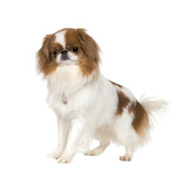
Japanese Chin
Lhasa apso
Pekingese
Pomeranian
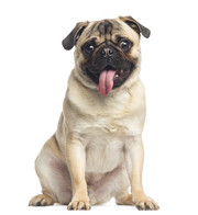
Pug
Shar-pei (Chinese shar-pei)
Shih Tzu
Staffordshire bull terrier


How does brachycephalic upper airway obstruction syndrome develop?
The syndrome comprises one or more of the following components:
Narrowing/stenosis of the external nares, obstruction of nasal vestibule by pronounced ventral alae, aberrant and hypertrophied nasal turbinates with increased mucosal contact point.
Nasopharyngeal narrowing and collapse.
Narrow pharyngeal dimensions, pharyngeal collapse, elongated soft palate and inflamed and extruded tonsils.
Narrow laryngeal dimensions, collapse of laryngeal cartilages, and redundant laryngeal soft tissues.
Hypoplastic trachea, especially in the bulldog and bronchial collapse (especially in the Pug).
Skull base malformation (e.g. medialization of the pterygoid processes).
What are the predisposing factors of brachycephalic upper airway obstruction syndrome?
Obesity.
Hot weather.
Exercise.
Excitement.
Stress.
Concurrent cardiac (heart) or pulmonary (respiratory) diseases.
Concurrent or secondary gastrointestinal diseases with gastro-oesophagal reflux.
What is the mechanism (Pathophysiology) of this syndrome?
Airflow through airways is impeded due to abnormal anatomy causing noisy breathing and the inability to take in sufficient oxygen to meet increased demands imposed by exercise. The restricted airflow leads to the increased inspiratory effort that causes increased negative pressure within the upper airways, leading to a high intrathoracic negative pressure which in turn leads to eversion of laryngeal tissues and airway collapse plus a sliding hiatal hernia of the stomach. The increased respiratory effort leads to upper airway oedema that further obstructs airflow. Regurgitation leads to acid reflux and increases pharyngeal and laryngeal inflammation thus, a vicious cycle is set in motion. Impeded airflow prevents adequate heat loss through panting, so animals rapidly become hyperthermic in hot weather, following exercise or during stress. If the respiratory vicious cycle is left untreated, the dog may develop pulmonary oedema, reduced arterial oxygen content, hypertension, and right-sided heart failure.
How is brachycephalic upper airway obstruction syndrome diagnosed?
History
Exercise intolerance.
Noisy breathing (snorting noise, ‘clicking sound’ when panting).
Loud snoring and disturbed sleeping. The dog may elevate the head when sleeping and/or hold a toy in the mouth to keep the mouth open while sleeping.
Collapse.
Signs may be exacerbated by exercise, excitement or hot weather.
Gagging/retching cough.
Regurgitation during exercise and/or excitement, or after eating/drinking
Clinical signs
Stenotic nares with restricted nasal flaring.
Stertorous or stridorous respiration increases laryngeal noise during laryngeal auscultation and inspiratory effort. Many of these signs only present after exercise. A short (3 minutes) trotting test (a.k.a. respiratory functional grading) is recommended.
Cyanosis (the tongue turns blue due to reduced oxygen in circulation).
Hyperthermia following exercise or stress due to the inability to cool body temperature by panting.
Laboratory investigation
Biochemistry:
Blood gas analysis -reduced arterial blood oxygen saturation in severe cases.
Whole-body barometric plethysmography (WBBP):
Whole-body barometric plethysmography (WBBP) is a non-invasive method that allows safe and repeated quantitative measurements of respiratory cycles on unsedated dogs.
WBBP flow trace shows a fixed-type obstruction, a dynamic-type obstruction, or both.
A BOAS index is used to calculate the respiratory patterns giving a severity score from 0 (BOAS free) to 100% (severe BOAS).
Endoscopic evaluation:
Oral examination and pharyngoscopy:
Pharyngeal narrowing.
Length of the soft palate.
Laryngoscopy
Laryngeal dimensions and degree of collapse of the larynx
Degree of oedema of the laryngeal mucosa.
Laryngeal granuloma.
Tracheobronchoscopy (Tracheoscopy):
Tracheal collapse
Bronchial collapse.
Rhinoscopy and nasopharyngoscopy:
Narrowing of the nasal vestibule (ventral alae).
Increased mucosal contact point.
Nasopharyngeal turbinate protrusion.
Nasopharyngeal cyst.
Diagnostic imaging
Thoracic and head radiography:
Hypoplastic trachea.
Aspiration pneumonia.
Severe hiatal hernia.

Computed tomography of the thorax, neck and head
Lower airway and the heart structure.
Nasopharyngeal dimension.
Swollen lateral nasal glands.
Soft palate length and thickness.
The thickness of the tongue.
Skull base abnormalities.
Hypoplastic and collapsed trachea and bronchi.
Stenotic nares and collapsed nasal vestibules.
Hiatal hernia.
Oesophagal diverticula/ redundant oesophagal mucosa.
How is brachycephalic upper airway obstruction syndrome treated?
a.) In case of an emergency:
The animal is sedated using either Acepromazine maleate (0.02-0.05 mg/kg) IV, IM or SQ, or Diazepam (0.2 mg/kg) IV may be combined with oxymorphone (0.5 mg/kg) or Butorphanol tartrate (0.3 mg/kg) IV, IM or SC.
A glucocorticoid (Prednisolone 0.5 mg/kg BID or dexamethasone 1-2 mg/kg IV) is administered to reduce laryngeal oedema.
Temporary tracheostomy may be required in severely cyanotic (blue) patients. A tracheostomy is an opening created at the front of the neck so that a tube can be inserted into the windpipe (trachea) to help an animal breathe. If necessary, the tube can be connected to an oxygen supply, and a breathing machine called a ventilator.
Endotracheal intubation may be considered during the crisis as an option.
Supplemental oxygen may be needed.
b) Removal exacerbating factors:
Cage rest to reduce stress and excitement.
Trazodone administration to reduce anxiety.
Cool patient if hyperthermic with fans, alcohol baths or cold water sprays.

c) Standard treatment:
Weight management (ideal body condition score 4-5/9).
Surgical correction of anatomical abnormalities:
Correction of stenotic nares enlargement and the enlarged and collapsed alar folds at nasal vestibules.
Correction of the oversized soft palate.
Correction of laryngeal collapse.
Removal of protruded tonsils.
Removal of the obstructive nasal turbinates (laser-assisted turbinectomy Laser-assisted turbinectomy (LATE)).
Medical management of gastrointestinal signs.
How is brachycephalic airway obstruction syndrome prevented?
Breeding should only be done with dogs that have been selected based on exercise tolerance tests (e.g. respiratory functional grading) and that have had airway evaluation by veterinary healthcare professionals to encourage group eradication.
References
Liu N-C, Sargan DR, Adams VJ, Ladlow JF (2015) Characterisation of Brachycephalic Obstructive Airway Syndrome in French Bulldogs Using Whole-Body Barometric Plethysmography. PLoS ONE 10(6): e0130741. https://doi.org/10.1371/journal.pone.0130741
MarchantI TW, DietschiI E, RytzI U, Schawalder P, Jagannathan V, Rasouliha SH, Gurtner C, Waldvogel AS, Harrington RS, Dro ̈gemu ̈ller M, KiddI K, Ostrander EA, Warr A, Watson M, Argyle D, Haar GT, Clements DN, Leeb T, Schoenebeck JJ (2019) An ADAMTS3 missense variant is associated with Norwich Terrier upper airway syndrome. PLoS Genetics doi.org/10.1371/journal.pgen.1008102.
Mielke B, Lam R & Ter Haar G (2017) Computed tomographic morphometry of tympanic bulla shape and position in brachycephalic and mesaticephalic dog breeds. Vet Radiol Ultrasound 58 (5), 552-558 PubMed.
Liu N-C, Oechtering G U, Adams V J et al (2017) Outcomes and prognostic factors of surgical treatments for brachycephalic obstructive airway syndrome in 3 breeds. Vet Surg 46 (2), 271-280 PubMed.
Heidenreich D, Gradner G, Kneissl S and Dupré G (2016) Nasopharyngeal dimensions from computed tomography of pugs and French bulldogs with brachycephalic airway syndrome. Vet Surg 45 (1), 83-90 PubMed.
Oechtering G U, Pohl S, Schlueter C et al (2016a) A novel approach to brachycephalic syndrome. 1. Evaluation of anatomical intranasal airway obstruction. Vet Surg 45 (2), 165-172 PubMed.
Oechtering GU, Pohl S, Schlueter C & Schuenemann R (2016b) A novel approach to brachycephalic syndrome. 2. Laser-assisted turbinectomy (LATE). Vet Surg 45 (2), 173-181 PubMed.
Pohl S, Roedler F S & Oechtering G U (2016) How does multilevel upper airway surgery influence the lives of dogs with severe brachycephaly? Results of a structured pre- and postoperative owner questionnaire. Vet J 210, 39-45 PubMed.
Kaye B M, Boroffka S A E B, Haagsman A N & ter Haar G (2015) Computed tomographic, radiographic and endoscopic tracheal dimensions in English bulldogs with grade 1 clinical signs of brachycephalic airway syndrome. Vet Radiol Ultrasound 56 (6), 609-616 PubMed.
Vilaplana Grosso F, Haar ter G & Boroffka S A E B (2015) Gender, weight, and age effects on prevalence of caudal aberrant nasal turbinates in clinically healthy English bulldogs: A computed tomographic study and classification. Vet Radiol Ultrasound 56 (6), 486-493 PubMed.
Schuenemann R & Oechtering G U (2014a) Inside the brachycephalic nose: intranasal mucosal contact points. JAAHA 50 (3), 149-158 PubMed.
Schuenemann R and Oechtering G (2014b) Inside the brachycephalic nose: conchal regrowth and mucosal contact points after laser-assisted turbinectomy. JAAHA 50 (4), 237-246 PubMed.
Oechtering G U (2010) Brachycephalic syndrome – new information on an old congenital disease. Veterinary Focus 20 (2), 2-9 VetMedResource.
Rooney N J (2009) The welfare of pedigree dogs: cause for concern. Journal of Veterinary Behaviour: Clinical Applications and Research 4 (5), 180–186 Journal of Veterinary Behaviour.
ter Haar G (2016a) Diseases of the nasal cavity and sinuses. In: Ear, Nose and Throat Diseases of the Dog and Cat. 1st edn, ed. R G Harvey & G ter Haar, pp 287-334. CRC Press, Taylor & Francis Group, London.
ter Haar G (2016b) Surgery of the nose. In: Ear, Nose and Throat Diseases of the Dog and Cat. 1st edn, ed. R G Harvey & G ter Haar, pp 449-474. CRC Press, Taylor & Francis Group, London.
Organisation(s)
Cambridge BOAS Research Group: www.vet.cam.ac.uk/boas.


Comments