


Addison's Disease in Pets: The Great Pretender of Veterinary Endocrinology

Updated: Aug 12
Introduction
Addison's disease is a rare but serious condition affecting pets, mainly dogs and sometimes cats, caused by insufficient hormone production from the adrenal glands. Early recognition and management can significantly improve your pet's quality of life.
If your dog seems "not right" for weeks—quiet, off food, with intermittent vomiting or diarrhoea—your vet might suggest testing for Addison's disease. Known as hypoadrenocorticism, it is called "the great pretender" because its symptoms mimic many other common illnesses, often leading to a delayed diagnosis until a medical emergency occurs. This article explains what Addison's disease is, its causes, diagnosis, and management, helping you understand your veterinary team's actions if the condition arises in your pet's care.

What Is Addison's Disease?

Addison's disease, also called hypoadrenocorticism, is a condition in which the adrenal glands fail to produce adequate amounts of the steroid hormones the body depends on for everyday physiological stability—chiefly cortisol and, in most cases, aldosterone (Merck Veterinary Manual, 2024). These hormones regulate important functions like stress response, blood pressure, and electrolyte balance. Without enough of these hormones, pets can experience a range of symptoms that often mimic other illnesses, making diagnosis tricky (Scott-Moncrieff, 2015). It is the mirror image of the much better-known Cushing's disease (hyperadrenocorticism), in which the adrenal glands produce too much cortisol.

Addison's disease can develop at any age but is most often diagnosed in middle-aged dogs, with females somewhat over-represented. Dogs are more commonly affected than cats, but both can develop the disease, and it has occasionally been reported in horses (Glebocka-Boag & Scudder, 2024).
Several breeds — including the Standard Poodle, West Highland White Terrier, Bearded Collie, Great Dane, Portuguese Water Dog, and Nova Scotia Duck Tolling Retriever — are disproportionately affected, suggesting a heritable component (Merck Veterinary Manual, 2024).
Standard Poodle West Highland Terrier Bearded Collie Great Dane Portuguese Water Dog Nova Scotia Duck Tolling Retriever
The Anatomy Behind the Disease

To comprehend Addison's disease, it is essential to understand the adrenal glands, which, despite their small size, play a crucial role. Each dog possesses two adrenal glands, located near each kidney. Each gland functions as two organs in one—comprising an inner medulla and an outer cortex.
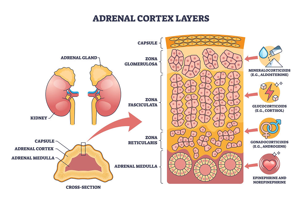
The cortex itself is divided into three concentric layers, each responsible for distinct hormonal functions (dvm360, 2026):
Zona glomerulosa (outermost) — the sole layer capable of producing aldosterone, the mineralocorticoid essential for maintaining salt and water balance.
Zona fasciculata (middle) — responsible for producing cortisol, the primary glucocorticoid.
Zona reticularis (innermost) — produces cortisol and precursors to sex steroids.

The medulla, located at the centre, produces catecholamines (epinephrine and norepinephrine) and is an entirely separate embryological structure from the cortex, which is why it typically remains unaffected even when the cortex is compromised. Figure 1 illustrates this layered anatomy and explains why Addison's disease typically results in the simultaneous impairment of both major cortical hormones.
The Physiology: One Gland, Two Failing Signals
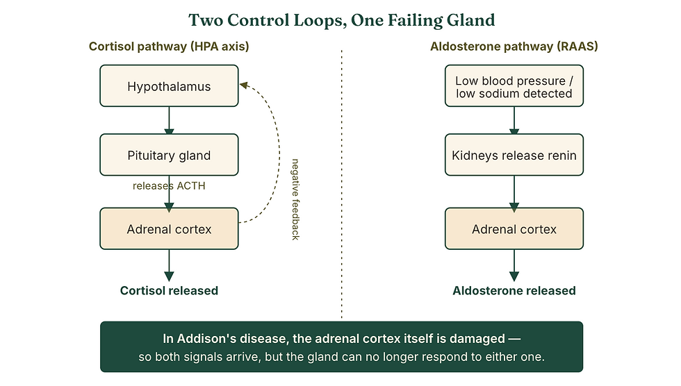
Two entirely separate hormone regulatory loops (axes) control cortisol and aldosterone, and Addison's disease typically silences both.

The HPA axis and cortisol.
The hypothalamus releases corticotropin-releasing hormone (CRH), which stimulates the pituitary gland to release adrenocorticotropic hormone (ACTH). ACTH travels through the bloodstream to the zona fasciculata, stimulating cortisol release. Cortisol, in turn, feeds back to suppress further CRH and ACTH release — a classic negative feedback loop. Cortisol is far more than a "stress hormone": it helps maintain blood glucose, vascular tone, and the integrity of the gastrointestinal lining, among many other functions.
The renin-angiotensin-aldosterone system (RAAS) and aldosterone.
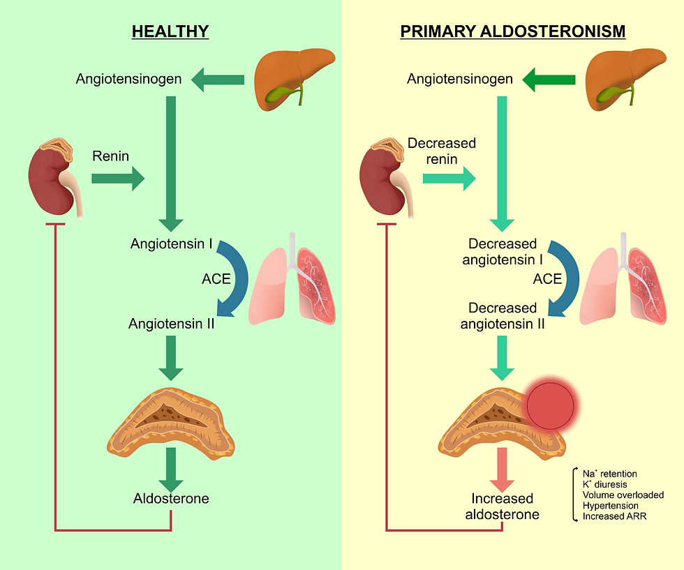
Aldosterone is regulated largely independently of ACTH. When the kidneys sense low blood pressure or low sodium, they release renin, which triggers a cascade ending in angiotensin II, which in turn stimulates the zona glomerulosa to release aldosterone. Aldosterone acts on the kidney to retain sodium and water while excreting potassium, which is essential for maintaining blood volume and pressure.
The Three Types of Adrenal Insufficiency (AI)
Addison's disease is categorised into three types based on the root cause and which part of the hormonal pathway fails:
1. Primary Hypoadrenocorticism (Typical & Atypical) - Type I AI
This is the most common form found in dogs. It originates directly within the adrenal glands themselves.
The Cause: In most dogs with naturally occurring Addison's disease, it is most commonly an immune-mediated condition where the dog’s own immune system mistakenly attacks and progressively destroys the entire adrenal cortex (outer layers of the adrenal glands) so both systems fail together—this is summarised and defined by cortisol deficiency combined with the sodium loss and potassium retention that come from aldosterone deficiency (Hughes-Hallett et al., 2025). The exact triggers remain incompletely understood, and a genetic predisposition has been identified through work on candidate susceptibility genes such as CTLA4 (Short et al., 2020). Other rare causes of primary disease include trauma, severe infections, adrenal tumours (neoplasia), localised bleeding, and granulomatous infiltration of the gland (Merck Veterinary Manual, 2024).
Typical Primary: Roughly 85% to 90% of the adrenal tissue must be destroyed before clinical symptoms appear. When this milestone is reached, the glands fail to produce both cortisol and aldosterone. This leads to severe electrolyte imbalances (dangerously high potassium and low sodium), which can cause acute collapse or heart arrhythmias.
Atypical Primary: Less commonly, in some instances, the immune destruction initially spares the aldosterone-producing layer, resulting in glucocorticoid deficiency with normal electrolytes – called "atypical" hypoadrenocorticism. The dog suffers from an isolated cortisol deficiency while maintaining normal electrolyte levels. However, atypical cases frequently progress into typical Addison's disease over time as tissue destruction continues.
Current veterinary endocrinology (hormone) terminology increasingly favours the more precise labels 'hyponatraemic/hyperkalaemic' and 'eunatraemic/eukalaemic' primary hypoadrenocorticism, respectively (Frontiers in Veterinary Science, 2025).

2. Secondary Hypoadrenocorticism - Type II AI
A second, distinct form of the disease is secondary hypoadrenocorticism, in which the adrenal insufficiency originates completely outside the adrenal glands, specifically inside the brain.
The Cause: This type occurs due to a structural defect, tumour, inflammation, or congenital issue in the pituitary gland. In this case, the pituitary gland fails to secrete adequate adrenocorticotropic hormone (ACTH) – either from pituitary disease or from abrupt withdrawal of long-term exogenous glucocorticoid therapy that has suppressed the patient's own HPA axis.
The Mechanism: The pituitary gland is responsible for secreting adrenocorticotropic hormone (ACTH), the chemical messenger that orders the adrenal glands to release cortisol. When the pituitary fails to produce ACTH, the adrenal glands completely stop producing cortisol. Because ACTH does not drive aldosterone production, dogs with secondary disease typically lose only cortisol, while their electrolytes and RAAS-driven aldosterone remain largely intact.
The Difference: Because aldosterone secretion is managed by the kidneys via the renin-angiotensin-aldosterone system (rather than pituitary ACTH), aldosterone production remains completely normal. Consequently, dogs with secondary Addison’s exhibit metabolic symptoms but do not display dangerous electrolyte imbalances.
This distinction matters clinically: It is essentially the same eunatraemic/eukalaemic ("atypical") presentation described above, just with a different underlying mechanism (primary cortical damage with early/mild aldosterone loss versus true secondary ACTH deficiency), and it is one reason electrolyte values alone cannot be used to rule the disease out (Guzmán Ramos et al., 2022; Merck Veterinary Manual, 2024).

3. Iatrogenic Hypoadrenocorticism
Iatrogenic conditions are inadvertently caused by medical intervention or prescription treatments.
The Cause: This most frequently occurs in two medical scenarios:
a) Abrupt Corticosteroid Withdrawal: If a dog is prescribed long-term, high-dose steroid medications (like prednisone) for allergies or immune diseases, the body assumes it has plenty of cortisol and shuts down its internal manufacturing from the zona fasciculata zone of the adrenal cortex. If the medication is stopped abruptly rather than tapered down gradually, the dormant adrenal glands cannot wake up fast enough to produce natural cortisol, throwing the dog into an immediate deficiency state.
b) Cushing's Disease Treatment: When a dog is being treated for Cushing's disease (an overproduction of cortisol), veterinarians use adrenal-suppressing drugs like mitotane or trilostane. If these medications are highly effective or given in a slightly high dosage, they can completely shut down or permanently damage the adrenal cortex, inducing accidental adrenal insufficiency.
Table I
Clinical Signs: Why It's Called "the Great Pretender"
The hallmark of Addison's disease is how unremarkable it can look for weeks or months before a crisis. Chronic, waxing-and-waning signs commonly include lethargy, poor appetite, vomiting, diarrhoea, weight loss, intermittent weakness, and — in some dogs — increased thirst and urination or bouts of shaking or trembling (Klein & Peterson, 2010). Because these signs overlap so heavily with gastrointestinal disease, kidney disease, and countless other conditions, dogs are often treated several times symptomatically before Addison's disease is ever suspected (Van Lanen & Sande, 2014).

The danger is that a stressful event — boarding, surgery, a concurrent illness, even a long car trip — can suddenly increase the body's demand for cortisol beyond what the failing adrenal glands can supply. This tips the patient into an acute Addisonian crisis: profound weakness, collapse, bradycardia (an unusually slow heart rate, in contrast to the fast heart rate expected with shock from other causes), hypothermia, weak pulses, and hypovolemic shock (Mitropoulou et al., 2022). Roughly 30% of dogs with hypoadrenocorticism are first recognised because of this kind of acute crisis (PMC, 2025). Electrolyte derangements — hyponatraemia, hyperkalaemia, hypochloraemia, and sometimes hypercalcaemia — are common and are responsible for many of the crisis signs, including the characteristic cardiac effects of high potassium (PMC, 2025).
How is Addison's disease detected?
Detecting Addison's disease early is crucial because symptoms can worsen quickly and lead to an Addisonian crisis, a life-threatening emergency.
a) Common clinical signs to watch for:
Lethargy and weakness: Pets may seem unusually tired or unwilling to move.
Loss of appetite and weight loss: A sudden drop in eating habits and weight can be a red flag.
Vomiting and diarrhoea: Digestive upset is common and may be mistaken for other illnesses.
Increased thirst and urination: Changes in drinking and bathroom habits can indicate a hormonal imbalance.
Shaking or trembling: This can be a sign of low blood sugar or electrolyte problems.
Collapse or seizures: In severe cases, pets may faint or have seizures due to electrolyte disturbances.
Figure IX traces this progression, from the earliest vague signs through to a diagnostic crisis and beyond.

These symptoms and clinical signs overlap with many other conditions; therefore, veterinarians perform specific blood tests to measure hormone levels and electrolytes, and sometimes, diagnosing Addison's disease depends on a combination of clues and, ultimately, a specific hormonal test.
b) Routine bloodwork clues.
Many affected dogs show a reduced sodium-to-potassium ratio (classically below about 27:1), azotemia (elevated kidney values, often from reduced blood flow to the kidneys rather than true kidney disease), mild anaemia, and — notably — the absence of a "stress leukogram," the pattern of white blood cell changes a sick, cortisol-producing body would normally show (Van Lanen & Sande, 2014). None of these findings is specific to Addison's disease on its own, which is why hormonal testing is required for a definitive diagnosis.
Baseline cortisol as a screening tool.
A single resting cortisol concentration above roughly 2 µg/dL (55 nmol/L) is highly effective at ruling Addison's disease out, since dogs with hypoadrenocorticism seldom have a baseline cortisol this high (Merck Veterinary Manual, 2024; American Animal Hospital Association [AAHA], 2023). However, a low baseline value alone cannot rule the disease in—many sick dogs without Addison's disease also have low baseline cortisol—so a low result must be followed by definitive testing (AAHA, 2023).
The ACTH stimulation test.

An ACTH Stimulation Test, which measures how the adrenal glands react to a synthetic trigger hormone, remains the gold-standard diagnostic test (Klein & Peterson, 2010). A baseline blood sample is drawn, synthetic ACTH (cosyntropin) is administered, and a second sample is drawn roughly one hour later. In a healthy dog, cortisol rises substantially in response to ACTH; in a dog with Addison's disease, the adrenal cortex is too damaged to respond, and cortisol remains low before and after stimulation — a "flatline" result (Merck Veterinary Manual, 2024). A post-ACTH cortisol concentration below the laboratory's diagnostic cutoff (commonly under 2 µg/dL) confirms the diagnosis (Merck Veterinary Manual, 2024; AAHA, 2023).
Veterinary endocrinologists continue to refine this protocol — recent large-scale data suggest that, once a full ACTH stimulation test is performed, the post-ACTH cortisol value alone may be sufficient for diagnosis, potentially allowing the baseline sample to be omitted in some settings to reduce cost without losing accuracy (Frontiers in Veterinary Science, 2025). The ACTH stimulation test is the most reliable method to confirm Addison's disease.
How is Addison's Disease in Dogs and Cats Managed?
Once diagnosed, Addison's disease requires lifelong management. The goal is to replace the missing hormones and keep your pet stable.
Here’s how management typically works:
a) Emergency stabilisation.
A dog presenting in an Addisonian crisis needs immediate intravenous crystalloid fluid therapy to correct hypovolaemia, shock, and dehydration — this alone is the single most important step, since restoring circulating volume also helps correct several electrolyte derangements (Merck Veterinary Manual, 2024). Alongside fluids, the emergency team typically addresses two additional threats:
Cardiac protection from hyperkalaemia.
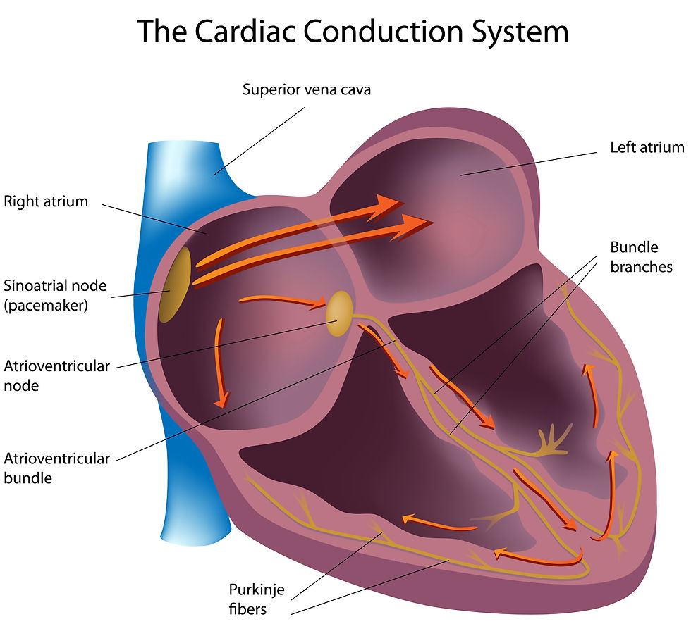
If severe hyperkalaemia is causing dangerous slowing of the heart rate or abnormal rhythms, 10% calcium gluconate can be given slowly by IV to stabilise heart muscle cells — this does not actually lower blood potassium but buys time while other treatments take effect (Merck Veterinary Manual, 2024). Dextrose, with or without a small dose of regular insulin, may also be given to help shift potassium back into cells (Merck Veterinary Manual, 2024).
Glucocorticoid replacement.

A fast-acting injectable corticosteroid is given as soon as IV access is secured. Dexamethasone is often favoured in the emergency setting specifically because, unlike hydrocortisone or prednisone, it does not interfere with the cortisol assay used in the ACTH stimulation test—allowing the diagnostic test to still be performed after treatment has started (Merck Veterinary Manual, 2024). Alternatively, some clinicians use a continuous infusion of hydrocortisone, which conveniently provides both glucocorticoid and mineralocorticoid activity during the acute crisis, though studies comparing the two approaches have not shown a clear advantage of one over the other (Mitropoulou et al., 2022). If hypoglycemia is also present, IV dextrose supplementation is added to the fluid plan (Merck Veterinary Manual, 2024).
b) Lifelong hormone replacement.
Treatment for primary types involves lifelong hormone replacement, while secondary or atypical variants generally require glucocorticoid replacement alone. With proper medical monitoring, dogs diagnosed with any form of adrenal insufficiency can live full, happy, and active lives. Once a patient is stable, treatment shifts to replacing the two missing hormone classes for life.
Mineralocorticoid replacement

Mineralocorticoid replacement is most often provided as desoxycorticosterone pivalate (DOCP), a long-acting injectable analogue of aldosterone given roughly every 3–4 weeks, or as oral fludrocortisone given once or twice daily (Merck Veterinary Manual, 2024). DOCP works by increasing sodium and water retention in the kidney while promoting potassium excretion, directly reversing the electrolyte disturbances of the disease (Bugbee et al., 2023). While the labelled DOCP dose is 2.2 mg/kg, a substantial body of evidence now supports starting at a lower dose (roughly 1.1–1.5 mg/kg) in most dogs, adjusting up or down based on follow-up electrolyte monitoring (AAHA, 2023).
Glucocorticoid replacement
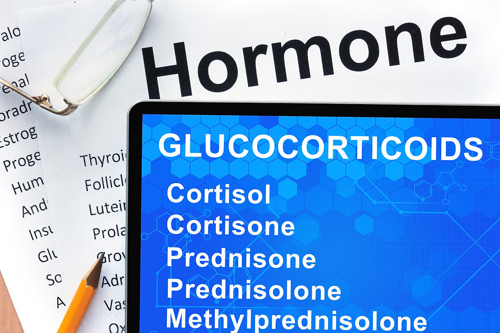
Glucocorticoid replacement is typically provided as low-dose oral prednisone, often at a lower dose than DOCP-treated dogs need during the initial crisis, since dogs receiving DOCP frequently require only modest supplemental glucocorticoid once stable (Merck Veterinary Manual, 2024).
c) Monitoring.
Regular check-ups

Frequent veterinary visits help catch any changes early. Electrolytes are rechecked roughly 10–14 days after each new DOCP dose and again near the end of the injection interval to fine-tune dosing; once a stable dose and interval are established, monitoring can typically be spaced out to every 3–6 months (AAHA, 2023). Dose requirements for fludrocortisone often rise over the years, whereas DOCP dosing tends to remain comparatively stable once established (Kintzer & Peterson, 2018).
Managing future stress

Because the adrenal glands can no longer mount their own stress response, owners are typically taught to increase the glucocorticoid dose temporarily during illness, surgery, travel, boarding, or other stressful events that can trigger an Addisonian crisis and to seek immediate veterinary attention for any signs of weakness, poor appetite, vomiting, or collapse (Van Lanen & Sande, 2014).
d) Diet and Lifestyle
Balanced diet
A nutritious diet supports overall health and helps maintain electrolyte balance.
Avoid stress
Minimise stressful situations that could upset your pet’s hormone balance.
Exercise
Moderate activity helps keep your pet fit, but avoid overexertion.

The Bottom Line for Owners
Addison's disease is a manageable, treatable condition—but only once it is recognised. With proper management, many dogs and cats with Addison's disease live happy, active lives. If your dog has recurring, unexplained bouts of lethargy, vomiting, diarrhoea, or poor appetite that seem to improve and then return, especially if there is ever an episode of sudden collapse, it is worth discussing hypoadrenocorticism with your veterinary team. A simple blood test can rule it out, and if it turns out to be the cause, most dogs go on to live full, active lives with a manageable injection or daily tablet routine. Owners often notice their pets regain energy and appetite once treatment starts.
Tips for Owners
Keep a medication schedule and set reminders.
Maintain regular vet visits for blood tests.
Educate family members and caregivers about the condition.
Prepare an emergency plan with your vet.
Consistency and vigilance are key to success.

Addison's disease may seem daunting, but with early detection and careful management, your pet can enjoy a good quality of life. If you notice any signs or have concerns, contact your veterinarian or The Andys Veterinary Clinic for advice and testing. Taking action early helps protect your furry friend from serious complications and keeps their tail wagging strong.
References
American Animal Hospital Association. (2023). Canine hypoadrenocorticism diagnostic testing and monitoring. In 2023 AAHA selected endocrinopathies of dogs and cats guidelines. Journal of the American Animal Hospital Association, 59(3), 113–135. https://www.aaha.org/resources/2023-aaha-selected-endocrinopathies-of-dogs-and-cats-guidelines/
American Animal Hospital Association. (2023). Canine hypoadrenocorticism therapy. In 2023 AAHA selected endocrinopathies of dogs and cats guidelines. Journal of the American Animal Hospital Association, 59(3), 113–135. https://www.aaha.org/resources/2023-aaha-selected-endocrinopathies-of-dogs-and-cats-guidelines/therapy-3/
Cornell University College of Veterinary Medicine. (n.d.). Addison's disease. Richard P. Riney Canine Health Centre. Retrieved July 22, 2026, from https://www.vet.cornell.edu/departments-centers-and-institutes/riney-canine-health-center/canine-health-information/addisons-disease
dvm360. (2026, July). Canine hypoadrenocorticism [Proceedings]. https://www.dvm360.com/view/canine-hypoadrenocorticism-proceedings
Frontiers in Veterinary Science. (2025). Reassessing the role of basal cortisol in ACTH stimulation testing for canine hypoadrenocorticism: Insights from a large UK referral and first-opinion dataset. Frontiers in Veterinary Science. https://www.frontiersin.org/journals/veterinary-science/articles/10.3389/fvets.2025.1686045/full
Galac, S. (2020). Hypoadrenocorticism in dogs and cats. In Clinical Handbook of Canine and Feline Endocrinology (pp. 145–162). Wiley Blackwell.
Glebocka-Boag, J., & Scudder, C. J. (2024). Hypoadrenocorticism in cats: A 40-year update. Journal of Feline Medicine and Surgery, 26(9).
Guzmán Ramos, P. J., Bennaim, M., Shiel, R. E., & Mooney, C. T. (2022). Diagnosis of canine spontaneous hypoadrenocorticism. Canine Medicine and Genetics, 9(1), Article 6. https://doi.org/10.1186/s40575-022-00119-4
Klein, S. C., & Peterson, M. E. (2009). Canine hypoadrenocorticism: Part I. Canadian Veterinary Journal, 50(1), 63–69.
Klein, S. C., & Peterson, M. E. (2010). Canine hypoadrenocorticism: Part II. Canadian Veterinary Journal, 51(2), 179–184.
Merck Veterinary Manual. (2024, July 11). Addison disease (hypoadrenocorticism) in animals. https://www.merckvetmanual.com/endocrine-system/the-adrenal-glands/addison-disease-hypoadrenocorticism-in-animals
Mitropoulou, A., Häuser, M. K., Lehmann, H., & Hazuchova, K. (2022). Comparison of hydrocortisone continuous rate infusion and prednisolone or dexamethasone administration for treatment of acute hypoadrenocortical (Addisonian) crisis in dogs. Frontiers in Veterinary Science, 8, Article 818515. https://doi.org/10.3389/fvets.2021.818515
Scott-Moncrieff, J. C. (2015). Hypoadrenocorticism. In E. C. Feldman, R. W. Nelson, C. E. Reusch, J. C. Scott-Moncrieff, & E. N. Behrend (Eds.), Canine and feline endocrinology (4th ed., pp. 485–520). Elsevier Saunders. https://www.scirp.org/reference/referencespapers?referenceid=2826902
Today's Veterinary Practice. (2022, March 30). Desoxycorticosterone pivalate for dogs with Addison's disease. https://todaysveterinarypractice.com/pharmacology/desoxycorticosterone-pivalate-for-dogs-with-addisons-disease/
Van Lanen, K., & Sande, A. (2014). Canine hypoadrenocorticism: Pathogenesis, diagnosis, and treatment. Topics in Companion Animal Medicine, 29(4), 88–95. https://doi.org/10.1053/j.tcam.2014.10.001
White, C. (2018). Use of desoxycorticosterone pivalate (DOCP) in the treatment of canine hypoadrenocorticism. Companion Animal, 23(2). https://doi.org/10.12968/coan.2018.23.2.82
This article is provided for general client education and does not replace individualised veterinary advice. If you believe your pet may be showing signs of Addison's disease, please contact The Andys Veterinary Clinic directly.


Comments