Aflatoxicosis: A Source of food poisoning in Dogs
- Dr Andrew Matole, BVetMed, MSc

- Jun 29, 2020
- 4 min read
Updated: Sep 22, 2024
What is aflatoxicosis?
Aflatoxicosis is a disease caused by ingesting highly poisonous fungal toxins in food or food scraps produced by a group of fungi called Aspergillus. These toxins, in a few micrograms per kilogram (µg/kg) concentrations, have adverse effects on both livestock and humans, with the liver being the principal organ affected [1]; [2] [3]. Four toxins out of the 17 aflatoxins are important in causing the disease in animals and humans. These are B1, B2, G1, and G2, with B1 being the most important [4]. The strains that produce this toxin are Aspergillus flavus and Aspergillus parasiticus on peanuts, soybeans, corn (maize), and other cereals either in the field or during storage when moisture content and temperatures are sufficiently high for mould to grow. Additional strains include M1 and M2. Acceptable and tolerable values range from 0.05 ppb to 0.5 ppb in different countries. The fungi grow as field fungi (infecting grains pre-harvest) and storage fungi (infecting grains and grain products in storage). Animals are poisoned by eating mouldy grain or grain products, e.g., mouldy bread [3].


Which animals are affected by Aflatoxins?
Aflatoxicosis is a serious health concern that impacts many animals, including growing poultry such as ducklings and turkey poults, young pigs, pregnant sows, calves, cats, dogs, cattle, sheep, and goats. This condition is caused by ingesting aflatoxins, toxic compounds produced by certain molds, particularly Aspergillus species.
Aflatoxiosis can lead to decreased growth rates, poor feed conversion, immunosuppression, and increased susceptibility to infections in poultry. Ducklings and turkey poults are particularly vulnerable due to their developing immune systems. Young pigs affected by aflatoxicosis may exhibit symptoms such as liver damage, reduced weight gain, and impaired reproductive performance in sows.
Pregnant sows exposed to aflatoxins are at risk of reproductive issues, including reduced fertility and increased rates of stillbirths or malformed piglets. Calves, as well as cats and dogs, can also suffer from aflatoxicosis, experiencing symptoms like liver damage, gastrointestinal disturbances, and even death in severe cases. [3].
Symptoms of aflatoxicosis in dogs and cats?
1. Anorexia
2. Weakness (lethargy),
3. Depression,
4. Vomiting,
5. Diarrhoea (melena, haematochezia),
6. Bleeding (haemorrhage or haemorrhagic diathesis),
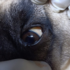
7. Icterus (jaundice)
8. Abdominal effusion (accumulation of fluid in the abdomen)
9. Peripheral oedema, and
10. Terminal encephalopathy [2].


How does aflatoxicosis develop in animals?
Among all animal species, dogs exhibit a particularly high sensitivity to aflatoxicosis, a condition characterized by the acute to subacute onset in these animals, typically lasting a few days. This acute manifestation in dogs starkly contrasts the chronic form of aflatoxicosis commonly observed in livestock. The process through which aflatoxins become toxic in dogs involves their conversion by cytochrome P450 enzymes within the liver cells. Subsequently, the highly reactive toxic elements, known as epoxides, bind to various cellular components such as DNA, proteins, and other macromolecules, causing significant cell damage. This destructive mechanism ultimately results in hepatocellular degeneration and necrosis, leading to impaired liver function. One notable consequence of this liver damage is the compromised synthesis of essential clotting factors, further exacerbating the health implications of aflatoxicosis in dogs.
How is aflatoxicosis detected or diagnosed?
1. Client history: access to mouldy dog food, mouldy food scraps, or use of mouldy bedding.
2. Clinical signs: Weakness, depression, vomiting, diarrhoea, melena (blood in stool), icterus.
3. Diagnostic investigation: food analysis and clinical pathology, including haematology, clinical chemistry, and urinalysis. Typical findings are elevated liver enzymes, hypoalbuminemia, and hyperbilirubinemia. In severe cases, results include prolonged bleeding times, thrombocytopenia, and elevations in bile acids [5].
4. Confirmation of diagnosis:
Discriminatory diagnostic features: History of exposure to mouldy food or bedding.
Definitive diagnostic features: demonstration of the mycotoxin in the food, stomach contents, or bedding is definitive.
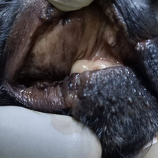
Gross autopsy findings: Icterus, widespread petechial and ecchymotic haemorrhage, ascites, subserosal oedema of the gallbladder. In severe cases, extensive haemorrhage and massive liver necrosis, while in longstanding cases, liver fibrosis [6].
Histopathology findings: Hepatocellular degeneration and necrosis, fatty change, megalocytosis, biliary hyperplasia, hepatic fibrosis. Renal proximal tubular necrosis may also be present [2].
How is aflatoxicosis treated?
Gastric decontamination may be relevant if ingestion is recent.
Remove the source of aflatoxins.
Cage rest
Avoid trauma
Offer a high quality, readily digestible diet, i.e., liver failure diet. The dog may require a special diet and care for the rest of its life
If icteric, protect the dog from UV light.
A blood transfusion may be required.
Depending on the extent of liver damage, Anabolic steroid therapy may enhance liver regeneration [5].
Fluid therapy
Vitamin K1
Antiemetics
Gastrointestinal protectants
NB: While aflatoxicosis is fatal in most patients with overt signs of intoxication, some dogs may recover slowly with long-term care.
How is aflatoxicosis prevented?
Do not feed table scraps or mouldy dog food to dogs.
Do not allow dogs to have access to garbage containing food scraps.
Do not use beddings for dogs that look or smell mouldy [6].
What causes treatment failure?
Substantial liver failure.
Fibrotic disrepair (cirrhosis) of the liver.
Fatal haemorrhage [8].
References
[1] C. Mutegi, P. Cotty and R. Bandyopadhyay, “Prevalence and mitigation of aflatoxins in Kenya (1960-to date),” World Mycotoxin Journal, vol. 11, no. 3, pp. 341 - 357, September 2018.
[2] R. Dalefield and P. Talcott, “Mycotoxicoses,” Vetstream Ltd, 2020. [Online]. Available: https://www.vetstream.com/treat/canis/diseases/mycotoxicoses. [Accessed 28 June 2020].
[3] G. D. Osweiler, “Aflatoxicosis.,” Merck Sharp & Dohme Corp, 2016. [Online].
[4] D. Dhanasekaran, S. Shanmugapriya, N. Thajuddin and A. Panneerselvam, “Aflatoxins and aflatoxicosis in human and animals.,” in Aflatoxins-Biochemistry and Molecular Biology, R. G. Guevara-Gonzalez, Ed., 2011, pp. 221-254.
[5] P. B., “Mycotoxins.,” The Veterinary clinics of North America. Small animal practice, vol. 32, no. 2, p. 409–419., 2002.
[6] Peterson and Talcott, Small Animal Toxicology., P. a. Talcott, Ed., WB Saunders Company, 2001, pp. 593-599.
[7] W. E. Huff, J. A. Doerr, P. B. Hamilton, D. D. Hamann, R. E. Peterson and A. Ciegler, “Evaluation of bone strength during aflatoxicosis and ochratoxicosis.,” Applied and environmental microbiology, vol. 40, no. 1, p. 102–107, 1980.
[8] Farlex Partner Medical Dictionary, “aflatoxicosis,” 22 June 2012. [Online]. Available: https://medical-dictionary.thefreedictionary.com/aflatoxicosis.
[9] G. D. Osweiler, in Toxicology., Philadelphia: Williams & Wilkins, 1996.
[10] K. H. Plumlee, Clinical veterinary toxicology., 6th ed., St. Louis, Mo: Mosby, 2004.


Comments