Can Pets Experience Urinary Incontinence? A Comprehensive Guide for Pet Owners
- Dr Andrew Matole, BVetMed, MSc

- Feb 25, 2024
- 14 min read
What is Urinary Incontinence?

Urinary incontinence (UI) in canines refers to the involuntary leakage or loss of urine from a dog's bladder, leading to unwanted urine leakage. It is a common problem in dogs and can manifest in various ways, including dribbling, puddles of urine left behind, or wetting their bedding or surroundings.
Dogs suffering from UI are typically unaware of or unable to control the passage of urine, leading to involuntary urination. This condition can vary in severity, from occasional small leaks to more significant and frequent episodes of urinary leakage.
The Structure and Anatomy of the Urinary Bladder
The structure and anatomy of the urinary bladder in humans and pets, such as dogs and cats, are quite similar in terms of function and basic structure, as both serve the same basic function of storing urine before it is expelled from the body. However, it's important to note that there can be variations in the size, shape, anatomy and function of the urinary bladder among different species of pets. For example, the shape and size of the bladder may vary in dogs, cats, and other animals with male dogs having a longer and more complex urethra than female dogs, making them more prone to urinary obstructions. Cats have a unique urinary system with their own set of characteristics and potential health issues.

Additionally, the mechanisms and reflexes controlling urination may differ among species. Understanding the specific anatomy and physiology of the urinary system in a particular pet species and breed is important for veterinary care and management of urinary issues as variations in anatomy and function impacts the susceptibility to certain urinary tract issues, such as urinary stones, infections, or bladder tumors, which may vary between species.
1. General Structure and Location:
The urinary bladder is a hollow, muscular organ located in the pelvic cavity. In humans, the urinary bladder is located in the pelvic cavity, just behind the pubic bone. In pets, the location of the bladder varies depending on the species. In dogs and cats, it is also situated in the pelvic cavity, similar to humans.
2. Shape and Size:

The shape of the urinary bladder can vary among individuals, but it typically has a rounded, sac-like shape. The size of the bladder can vary significantly between species and even among individuals within a species. For example, a human bladder can typically hold around 400-600 milliliters of urine, while the bladder in pets like dogs and cats may be smaller and hold less urine in proportion to their body size. In dogs and cats, the size and shape can vary based on the breed and size of the animal.
3. Muscular Wall and Internal Structures:

The urinary badder is made up of several layers of tissue, including mucosa, submucosa, muscularis, and serosa. The outer most layer surrounding the bladder, also called the outer connective tissue layer or adventitia or serosa, anchors the bladder to surrounding structures providing structural support. The muscularis layer , also called the detrusor muscle, of the urinary bladder contains smooth muscle fibers that contract to expel urine from the bladder.

The contraction of the muscular wall is under voluntary control in humans, allowing us to control when we empty our bladders. In most pets, it is under involuntary control. The submucosa, a layer of connective tissue that supports the mucosal layer, contains blood vessels and nerves that regulate bladder function. The innermost layer, called the mucosa, is lined with specialized cells known as transitional epithelium, which can stretch to accommodate urine without leaking.
4. Nerves and Blood Supply:

Both humans and pets have a complex network of nerves and blood vessels that supply the urinary bladder. These nerves are responsible for signaling when it's time to empty the bladder.

The urinary bladder receives its blood supply from various arteries, including the superior and inferior vesical arteries plus branches of the internal iliac artery in both humans and animals. Nerves, including the autonomic nervous system, play a crucial role in controlling bladder function. The bladder receives nerve signals from the autonomic nervous system, controlling involuntary contractions for urination. These nerves regulate the contraction of the detrusor muscle and the relaxation of the internal and external sphincters to control urination.
5. Urethra Connection:

The bladder is connected to the external environment through the urethra, which is a tube that carries urine from the bladder to the external urinary meatus (opening). The length and structure of the urethra can vary among species, with males typically having longer urethras than females.
6. Sphincters:
Both humans and pets have sphincters, which are muscles that control the flow of urine in and out of the bladder. There are two main types:
i) Internal Sphincter: Involuntary smooth muscle located at the bladder neck, which relaxes during urination.
ii) External Sphincter: Voluntary skeletal muscle that surrounds the urethra and allows for conscious control over urination.
7. Supportive Structures:
In humans, the bladder is supported by ligaments and the pelvic floor muscles.
In pets, the bladder is also supported by surrounding various structures and connective tissues that help maintain its position and functionality. These supportive structures are crucial for the proper functioning of the bladder. The supportive structures of the urinary bladder in both pets (such as dogs and cats) and humans include:
i). Pelvic Floor Muscles: The pelvic floor muscles play a significant role in supporting the urinary bladder in both humans and pets. These muscles form a supportive hammock-like structure that helps maintain the position of the bladder within the pelvic cavity.
ii). Ligaments: Ligaments provide additional support to the bladder by attaching it to surrounding structures. In humans, the pubovesical ligament and the puboprostatic ligament (in males) or the pubourethral ligament (in females) help anchor the bladder in place. In pets, similar ligaments may exist, depending on the species.
iii). Connective Tissues: The bladder is surrounded by connective tissues, including fascia and other supporting structures, which help maintain its structural integrity and position within the pelvic cavity.

iv). Peritoneum: The urinary bladder is partially covered by a serous membrane called the peritoneum. The peritoneum helps to support and stabilize the bladder within the abdominal cavity.
v). Adventitia or Serosa: The outermost layer of the bladder wall is composed of connective tissue known as adventitia or serosa. This layer provides additional support and helps anchor the bladder to surrounding structures.
vi). Pelvic Bones: The bony pelvis provides a stable foundation for the urinary bladder. The bladder is situated within the pelvic cavity and is supported by the pelvic bones, contributing to its overall stability.
vii). Urogenital Diaphragm: In both humans and some animals, including dogs, the urogenital diaphragm is a muscular structure that provides support to the bladder and other urogenital organs. This diaphragm is formed by the external urethral sphincter muscle and associated connective tissues.
These supportive structures work together to maintain the position and function of the urinary bladder. The pelvic floor muscles, ligaments, connective tissues, and bony structures collaborate to ensure that the bladder can adequately store urine and release it when necessary. Dysfunction or weakness in these supportive structures can contribute to conditions such as urinary incontinence or pelvic organ prolapse. Understanding the anatomy and function of these supportive structures is essential for the diagnosis and treatment of bladder-related issues in both humans and pets.
What is the basic Function of the urinary bladder?
The primary function of the urinary bladder in both humans and pets is to store urine produced by the kidneys until it is ready to be eliminated from the body. When the bladder fills with urine, stretch receptors signal the nervous system, and when it's appropriate, the bladder contracts to expel urine through the urethra.

While the basic structure and function of the urinary bladder are similar in both humans and pets, there can be variations in size, shape, and specific anatomical features depending on the species. Additionally, the control of bladder function may differ, with humans having more voluntary control over urination compared to most pets.
What causes Urinary Incontinence (UI) in Pets?
Urinary incontinence, often referred to as UI, is a medical condition characterized by the involuntary leakage or loss of urine from a dog's bladder in dogs of any age, breed, or gender, but it is more commonly observed in certain groups or conditions, such as:
1. Spayed Female Dogs: Female dogs that have been spayed (neutered) are more prone to develop urinary incontinence, especially as they age. This condition is often associated with a weakening of the urethral sphincter muscle. Urethral Sphincter Mechanism Incompetence (USMI) is more frequently seen in spayed female dogs but can also affect males and intact females. It often develops several years after spaying and can result from changes in hormonal levels, leading to a weakening of the muscles that control the closure of the urethra.

2. Larger Breeds: Some large dog breeds, such as Doberman Pinschers, Boxers, and Old English Sheepdogs, are more susceptible to urinary incontinence.


3. Aging Dogs: As dogs get older, their muscles and tissues can weaken, including those responsible for maintaining bladder control. This can lead to an increased risk of incontinence.

4. Medical Conditions: Certain medical conditions, such as urinary tract infections (UTIs), bladder stones, and neurological disorders, can also contribute to urinary incontinence in dogs.


5. Congenital (anatomical) Abnormalities: In some cases, urinary incontinence may be due to congenital abnormalities, such as ectopic ureters, which can lead to improper urine flow.
6. Other potential causes of UI in dogs include behavioural problems. Behavioral issues leading to incontinence are less common than medical causes, but they can still contribute to urinary problems. Some of the behavioral problems that may lead to incontinence in dogs include:

i) Excitement Urination: Some dogs may urinate involuntarily when they are excited or anxious. This is more common in puppies and young dogs and often occurs in response to new people, visitors, or during play. It is generally not a medical issue but a behavioral one.

ii) Submission or Submissive Urination: Dogs may urinate as a submissive gesture, especially when they feel intimidated or in the presence of dominant individuals. This is more common in shy or anxious dogs and can be triggered by specific situations or interactions.

iii) Marking Behaviour: Marking is a normal behaviour in which dogs deposit small amounts of urine to establish territory or communicate with other dogs. However, inappropriate marking indoors can lead to accidents and incontinence issues.

iv) Separation Anxiety: Dogs with separation anxiety may exhibit various behaviours, including inappropriate urination. When left alone, anxious dogs may urinate as a response to stress or fear. This is different from true incontinence, as it is related to emotional distress.

v) Territorial Issues: Some dogs may urinate indoors as a way of marking their territory. This behavior is more common in unneutered males, but spayed females and neutered males may also exhibit territorial marking.
Accurate diagnosis and identification of the underlying cause are essential to determine the most appropriate treatment for the affected dog.
When Does Urinary Incontinence Typically Begin?
Urinary Incontinence typically develops in young or middle-aged dogs.
Onset in older dogs requires further diagnostic investigation for underlying causes.
Addressing the underlying reason for polyuria (excessive urination) may improve Urinary Incontinence.
What is the mechanism of Urinary Incontinence?
Urinary incontinence is a complex condition characterized by the involuntary loss of urine due to the inability to control bladder function. The mechanism behind urinary incontinence development is multifactorial and there are various underlying mechanisms depending on the specific type of incontinence. The mechanisms involved include hormonal changes, anatomic factors, and tissue characteristics. Hormonal changes may affect smooth muscle contractility in the lower urinary tract, e.g., oestrogen decline after neutering may affect the vasculature and supporting tissues of the urethra Bladder position also plays a role, e.g., pelvic bladder can lead to pressure gradient and leakage while reduction in adrenergic α-receptors is often targeted for treatment
There are various types of urinary incontinence, and their mechanisms differ. The main types include stress incontinence, urge incontinence, overflow incontinence, functional incontinence, mixed incontinence, Hormonal Influence (Estrogen Deficiency), Neurological Disorders, Urinary Tract Infections (UTIs), and Bladder Stones or Tumors:
1. Stress Incontinence
Mechanism: Stress incontinence is often caused by weakened pelvic floor muscles and

sphincters, which support the bladder and control the release of urine. Factors such as pregnancy, whelping, obesity, and aging can contribute to the weakening of these supportive structures, leading to the inability to withstand pressure on the bladder during activities like coughing, sneezing, or exercising when pressure is exerted on the bladder and the weakened muscles may not provide sufficient support, resulting in urine leakage.
2. Urge Incontinence
Mechanism: Urge incontinence is usually associated with an overactive bladder muscle,

leading to sudden, intense urges to urinate. The bladder muscle contracts involuntarily, causing the urgency to urinate even when the bladder is not full resulting in leakage. The detrusor muscle contracts involuntarily, causing a sudden and intense urge to urinate. This can be caused by neurological issues, bladder irritation, or other factors that disrupt normal bladder function.
3. Overflow Incontinence

Mechanism: This occurs when the bladder does not empty properly, causing it to become overly full and leading to leakage. Causes include obstruction of the urethra (e.g., due to an enlarged prostate in men and male dogs), weak detrusor muscle contractions, or neurological conditions affecting bladder emptying.
4. Functional Incontinence
Mechanism: Functional incontinence is not directly related to bladder dysfunction or urethra but is instead due to physical or cognitive impairments that hinder an individual's or a pets ability to reach the toilet or toilet area in time. This can include conditions such as arthritis, dementia, or mobility issues.
5. Mixed Incontinence
Mechanism: This type involves a combination of more than one form of urinary incontinence. For example, an individual or pet may experience a combination of stress and urge incontinence or other types of incontinence simultaneously. The mechanisms are a combination of those associated with each specific type.
6. Hormonal Influence (Oestrogen Deficiency)

Mechanism (applicable to canine urinary incontinence): In spayed female dogs, a decrease in oestrogen levels can lead to weakened urethral sphincter tone, contributing to incontinence. Onset usually occurs within 3 to 4 years of neutering.
7. Neurological Disorders

Mechanism: Neurological conditions or injuries affecting the nerves controlling the bladder and sphincter muscles can disrupt normal signaling and lead to incontinence.
8. Urinary Tract Infections (UTIs)

Mechanism: UTIs can cause irritation and inflammation of the bladder, leading to urgency and involuntary contractions that result in leakage.
9. Bladder Stones or Tumours

Mechanism: Obstructions caused by bladder stones or tumors can interfere with normal urinary flow, triggering incontinence.
It's important to note that urinary incontinence can be a symptom of an underlying medical condition, and its mechanism may vary based on the specific cause
How is Urinary Incontinence (UI) Diagnosed?
1. History

The veterinarian gathers detailed information about the dog's medical history from the owner, including the onset of incontinence, any recent changes in behaviour, any potential triggering events, notes any medications the dog is currently taking and inquires about the dog's spaying/neutering status plus details about the dog's general health.
2. Physical Examination

A comprehensive physical examination is usually conducted to check for any abnormalities, pain, or signs of systemic diseases. This may include:
Rectal examination with urethra palpation can help diagnose obstructive processes (e.g., urethral neoplasia, prostatic hyperplasia).
Observing the dog's posture during urination especially in males, is essential to detect functional obstruction, to identify any abnormalities in the stream or posture, signs of discomfort and overflow incontinence, ruling out strictures, uroliths, and extramural compressive lesions..
Measuring residual urine volume is necessary in dogs with narrow urine streams, stranguria, or urine dripping.
Neurologic and orthopedic evaluation should be performed if the dog cannot posture normally for urination.
3. Urinalysis

A urinalysis with sediment examination is a crucial diagnostic tool. It helps identify any signs of urinary tract infection (UTI), bladder stones, or other urinary abnormalities. The specific gravity of the urine should be noted, as a low specific gravity may indicate underlying issues.
4. Blood-work

Blood tests are recommended, especially if the dog has a low urine-specific gravity. Blood-work can provide additional information about the dog's overall health and help identify potential systemic issues.
5. Neurological Examination

A neurological evaluation should be performed if the dog shows signs of difficulty posturing normally for urination. This helps assess nerve function related to bladder control, as neurological issues can contribute to incomplete emptying of the bladder.
6. Imaging Studies

Imaging studies such as abdominal ultrasound or contrast radiography may be conducted to visualize and evaluate the lower urinary tract, identify any structural (anatomical) abnormalities, or rule out bladder stones.
7. Cystoscopy

In some cases, a veterinarian may perform cystoscopy, a procedure that involves inserting a thin tube with a camera into the bladder to directly visualize the interior of the bladder and urethra, especially in cases where medical therapy is not effective, and identify any abnormalities.
8. Urodynamic Testing

In certain cases, urodynamic testing may be necessary to assess bladder function and rule out overactive bladder as a cause of incontinence, for example, in cases where overactive bladder or detrusor hyper-reflexia is suspected.
The Urodynamic Test
Urodynamic testing is a diagnostic procedure used to assess the function of the urinary system, particularly the bladder and urethra. It provides valuable information about how these structures store and release urine. While more commonly performed in humans, urodynamic testing is sometimes used in veterinary medicine, particularly when investigating urinary incontinence or bladder dysfunction. The procedure typically involves the following steps:
1. Patient Preparation:
The animal may need to be sedated or anesthetized, depending on its temperament and the testing requirements. This ensures the animal remains still during the procedure.
2. Catheterization:
A catheter is inserted into the bladder through the urethra. In some cases, a small catheter may also be inserted into the rectum to measure intra-abdominal pressure.
3. Filling the Bladder:
The bladder is filled with a sterile saline solution through the catheter. The rate at which the bladder is filled and the volume of the solution are carefully controlled during this phase.
4. Measurement of Pressures:
Special sensors on the catheters measure pressures within the bladder, urethra, and possibly the rectum. This helps evaluate how well the bladder stores urine and the pressure exerted during filling.
5. Observation of Responses:
The veterinarian may observe the animal's responses to bladder filling, including any involuntary contractions or spasms of the bladder muscle.
6. Voiding Phase:
Once the bladder is adequately filled, the animal may be allowed to void or expel urine. The pressures during voiding are also measured.
7. Analysis of Results:
The data collected during urodynamic testing are analyzed to assess bladder function, urethral resistance, and other factors influencing urinary continence.
Urodynamic testing helps veterinarians understand the underlying causes of urinary issues, such as incontinence or abnormal voiding patterns. It can provide insights into conditions like detrusor overactivity, urethral sphincter mechanism incompetence, or functional obstruction. It's important to note that while urodynamic testing can be valuable, it is not routinely performed in all cases of urinary incontinence in animals. The decision to use urodynamic testing is based on the specific clinical presentation, the results of other diagnostic tests, and the veterinarian's judgment.
How is Urinary Incontinence (UI) Treated in Dogs?
Urinary incontinence (UI) can be a distressing condition for both dogs and their owners, but various medical and surgical treatment options are available to manage and improve the condition, depending on the specific cause and severity of the incontinence. It's essential to consult with a veterinarian if you suspect your dog is experiencing urinary incontinence to receive a proper diagnosis and develop a suitable treatment plan to improve your pet's quality of life.
Treatment for urinary incontinence in dogs depends on the underlying cause. It may include medications to strengthen the urethral sphincter, hormone therapy (especially for spayed females), treatment of underlying medical conditions, and in some cases, surgical interventions.
Initial treatment includes α-agonist medications and oestrogen compounds. Combination therapy may be beneficial in some cases, however, monitoring blood pressure is important for α-agonist therapy. Surgical interventions, such as artificial urethral sphincter placement or urethral collagen injections, may be considered if medical therapy fails.
What are alpha agonist medications?
Alpha-agonist medications are drugs that stimulate alpha-adrenergic receptors in the body. These receptors are part of the sympathetic nervous system, which is responsible for the "fight or flight" response. Alpha-adrenergic receptors are further classified into two subtypes: alpha-1 receptors and alpha-2 receptors.
1. Alpha-1 Agonists:
These drugs stimulate alpha-1 receptors, leading to various physiological effects. Activation of alpha-1 receptors generally causes smooth muscle contraction in organs such as blood vessels, the bladder, and the prostate. Medications that act as alpha-1 agonists are often used in conditions such as:
Hypertension: By causing vasoconstriction (narrowing of blood vessels), these drugs can increase blood pressure.
Benign Prostatic Hyperplasia (BPH): Constriction of alpha-1 receptors in the prostate can relieve symptoms associated with BPH.
2. Alpha-2 Agonists:
These drugs stimulate alpha-2 receptors, leading to a decrease in the release of norepinephrine and other neurotransmitters. This results in a reduction of sympathetic nervous system activity.
Medications that act as alpha-2 agonists are used in various conditions, including:
Hypertension: By reducing sympathetic outflow, these drugs can lower blood pressure.
Attention Deficit Hyperactivity Disorder (ADHD): Some alpha-2 agonists are used to manage symptoms in ADHD.
Anaesthesia: Alpha-2 agonists can be used as sedatives and to reduce the requirement for other anesthetics.
Examples of medications that act as alpha-agonists include:
Alpha-1 Agonists:
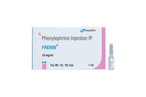
- Phenylephrine
- Pseudoephedrine
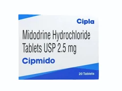
- Midodrine


Alpha-2 Agonists:
- Clonidine
- Methyldopa


It's important to note that these medications should be used under the supervision of a healthcare professional, as they can have side effects and may not be suitable for everyone.
Overactive Bladder
Overactive bladder may be a cause in some cases and can be treated with antimuscarinic drugs. Urodynamic testing may be needed for definitive diagnosis.

Functional Urethral Obstruction
In some cases, male dogs with poor response to traditional Urethral Sphincter Mechanism Incompetence (USMI) therapy may have functional obstruction instead.
This involves an inability to empty the bladder, not weak urethral sphincters.
Medical Treatment
Muscle relaxation and anxiolytic therapy are common approaches.
Smooth and skeletal muscle relaxants may be required.
Anxiolytic medications like trazodone or fluoxetine may help stressed or anxious dogs.
Urethral catheterization instructions for dog owners can provide temporary relief.
Long-term Outcomes
Limited information is available on long-term outcomes.
Some dogs may require significant dose adjustments to determine optimal therapy.
Urethral stenting is a salvage procedure for non-responders but may have significant adverse effects.
Conclusion
The mechanisms behind UI in dogs are complex and not fully understood.
Treatment depends on whether it involves holding urine during bladder filling or emptying the bladder during urination.
Medical therapy targets urethral muscle function, while surgical and cystoscopic interventions are options if medical therapy fails.
References:
Byron JK, Taylor KH, Phillips GS, Stahl MD. Urethral sphincter mechanism incompetence in 163 neutered female dogs: diagnosis, treatment, and relationship of weight and age at neuter to development of disease. J Vet Intern Med.2017;31(2):442-448. doi: 10.1111/jvim.14678.
Forsee KM, Davis G, Mouat EE, Salmeri KR, Bastian RP. Evaluation of the prevalence of urinary incontinence in spayed female dogs: 566 cases (2003-2008). J Am Vet Med Assoc. 2013;242(7):959-62. doi: 10.2460/javma.242.7.959.
Thrusfeld MV, Holt PE, Muirhead RH. Acquired urinary incontinence in bitches: its incidence and relationship to neutering practices. J Small Anim Pract. 1998;39:559-566.
Reichler IM, Hung E, Jöchle W, et al. FSH and LH plasma levels in bitches with differences in risk for urinary incontinence. Theriogenology. 2005;63:2164-2180.
Reichler IM, Jöchle W, Piché CA, Roos M, Arnold S. Effect of a long acting GnRH analogue or placebo on plasma LH/FSH, urethral pressure profiles and clinical signs of urinary incontinence due to sphincter mechanism incompetence in bitches. Theriogenology. 2006;66:1227-1236.
Adams WM, DiBartola SP. Radiographic and clinical features of pelvic bladder in the dog. J Am Vet Med Assoc. 1983;182(11):1212-1217.
Acierno MJ, Labato MA. Canine incontinence. Compend Contin Educ Pract Vet. 2006;28:591-598.
Holt P. Urinary incontinence. In: Holt P (Ed): Urological Disorders of the Dog and Cat: Investigation, Diagnosis and Treatment. London, UK: Manson Publishing; 2008:134-159.
Reeves L, Adin C, McLoughlin M, Ham K, Chew D. Outcome after placement of an artificial urethral sphincter in 27 dogs. Vet Surg. 2013;42(1):12-18. doi: 10.1111/j.1532-950X.2012.01043.x.
Barth A, Reichler IM, Hubler M, Hässig M, Arnold S. Evaluation of long-term effects of endoscopic injection of collagen into the urethral submucosa for treatment of urethral sphincter incompetence in female dogs: 40 cases (1993-2000). J Am Vet Med Assoc. 2005;226(1):73-76.
Byron JK, Chew DJ, McLoughlin ML. Retrospective evaluation of urethral bovine cross-linked collagen implantation for treatment of urinary incontinence in female dogs. J Vet Intern Med. 2011;25(5):980-984.
Rawlings C, Barsanti JA, Mahaffey MB, Bement S. Evaluation of colposuspension for treatment of incontinence in spayed female dogs. J Am Vet Med Assoc. 2001;219(6):770-775.
Aaron A, Eggleton E, Power C, Holt PE. Urethral sphincter mechanism incompetence in male dogs: a retrospective analysis of 54 cases. Vet Rec. 1996;139:542-546.
Palerme JS, Mazepa A, Hutchins RG, Ziglioli V, Vaden SL. Clinical response and side effects associated with testosterone cypionate for urinary incontinence in male dogs. J Am Anim Hosp Assoc. 2017;53(5):285-290.
Plumb DC. Plumbs Veterinary Drug Hanbdbook, 9th ed. Ames, IA: Wiley-Blackwell; 2018.
Lane IF, Westropp JL. Urinary incontinence and micturition disorders: pharmacologic management. In Kirk’s Current Veterinary Therapy, 14th ed. St. Louis, MO: Elsevier, 2009;955-959.


Comments