What is Cherry Eye?
- Dr Andrew Matole, BVetMed, MSc
- Sep 14, 2023
- 4 min read

Cherry eye in dogs is a common term for a condition known as prolapsed nictitating membrane, nictitans gland prolapse, prolapsed third eyelid gland,or prolapse of the third eyelid gland.
It is a condition where the gland of the third eyelid becomes swollen and protrudes from the inner corner of the eye, giving it a cherry-like appearance. It can happen in one or both eyes at the same time.
How is the anatomy of the nictitans gland?

Dogs have a third eyelid, also known as the nictitating membrane or haw, which contains a tear-producing gland called the nictitans gland or the lacrimal gland of the third eyelid. This gland helps in maintaining the eye's moisture and health. The nictitating membrane or third eyelid is located in the inner corner of each eye.
Why is the nictitans gland Significant?

The gland produces up to 70% of the aqueous phase of pre-corneal tear film. Prolapse and exposure induce adenitis (inflammation of the gland), potentially decreasing tear production. Ocular surface irritation may result from physical rubbing, or low tear production.
What is the predisposition of Cherry Eye in dogs?
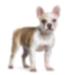
The condition is predisposed to young dogs aged between 6 months and 2 years old. It primarily affects certain breeds and species, including:-
Bulldogs,
Cocker Spaniels,
Shih Tzus,
Great Danes
Lhasa Apsos
American Cocker Spaniels
Pekingese
Beagles
Mastiffs


What is the cause of Cherry Eye?

The exact cause of cherry eye is not fully understood, but it is believed to occur when the connective tissue that normally holds the gland in place weakens or becomes damaged. Genetics may play a role in predisposing certain breeds to this condition. As a result, the gland protrudes or "prolapses" from its normal position and becomes visible as a reddish-pink mass in the corner of the eye. This gives it the appearance of a cherry, hence the name "cherry eye."
What are the clinical signs of Cherry Eye?
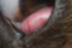
The most obvious symptom is the protrusion of a red or pink mass at the inner corner of the eye, visibly seen as a red or pink mass. Dogs with cherry eye may also exhibit signs of eye discomfort, such as squinting, excessive tear production, irritation in the affected eye or rubbing at the affected eye. Generally does not cause corneal ulceration, but in severe cases may inhibit healing of coinciding corneal ulcer or other eye problems. Acute cases may return to normal position with manipulation but longer-standing cases remain prolapsed.
How is Cherry Eye treated?
Cherry eye should be treated promptly to prevent complications. The best treatment for cherry eye in dogs depends on several factors including the specific case, the severity of the condition, the dog's age, the dog's overall health, the extent of the prolapse and the veterinarian's assessment. There is no one-size-fits-all approach, but here is an overview of the treatment options commonly considered. Treatment options include:
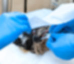
Surgical replacement (Repositioning): This is the most common treatment.
Description: This is often the preferred treatment method for cherry eye. It involves surgically repositioning the prolapsed gland back to its normal position.
Procedure: The veterinarian makes an incision near the cherry eye and carefully returns the gland to its proper location and sutures are used to secure the gland and reinforcing the connective tissue to promote healing and prevent recurrence. There always remains a chance of recurrence.
Advantages: Surgical repositioning aims to maintain the eye's natural tear production and lubrication, reducing the risk of dry eye. Surgical replacement typically provides the best chance for long-term resolution of the issue while preserving the gland's role in tear production and eye health.
Gland removal (Partial or Complete Removal):
Description: Only in certain cases, veterinarians may opt to remove the prolapsed gland partially or completely where the gland is severely damaged, non-functional, or if other treatments have failed, removal of the gland may be considered. However, this is generally not the first choice because it can lead to decreased tear production and potential long-term eye dryness (keratoconjunctivitis sicca or dry eye).
Advantages: This procedure eliminates the risk of further cherry eye episodes but may result in decreased tear production, potentially leading to dry eye.
Considerations: Dry eye can be managed with medication if necessary after the gland removal.
Gland Tacking (Alternative Approach): Gland tacking involves securing the gland in place within the third eyelid. This technique aims to prevent future prolapse while retaining the gland's function.
Advantages: Gland tacking can be a viable alternative when surgical replacement is not an option. It helps prevent recurrence while maintaining tear production. It is a surgical technique that secures the gland in place to prevent future prolapse.
4. Medical Management: Initially, symptomatic treatment can be done through topical ocular lubricants or topical ocular antibiotics/anti-inflammatories if there is significant adenitis (inflammation of the nictitating gland). In some cases, veterinarians may attempt to reduce the prolapse using topical medications and massage. However, this is often less effective, and surgery is eventually required
What are the complications if left untreated?
If left untreated, cherry eye can lead to more serious eye problems, such as dry eye or .
If left untreated, cherry eye can lead to eye dryness (keratoconjunctivitis sicca), irritation (conjunctivitis), and corneal ulcers, which can be painful and require more extensive treatment.
How is Cherry Eye treatment Monitored?
Additionally, post-operative care and follow-up visits are essential to monitor the dog's progress and ensure the surgical site heals properly, taking into account the specific circumstances of each case. The following are essential to observe hen monitoring:-
Ensure no corneal ulceration due to rubbing of sutures in first days post-operatively.
Prophylactic topical ocular antibiotics/anti-inflammatory medication for 7-10 days post-operatively.
Oral anti-inflammatories if patient exhibits discomfort.
How is Cherry eye Prevented?
There are no known methods to prevent cherry eye, as it is often linked to genetics. However, responsible breeding practices may help reduce the risk in susceptible breeds.

References
Stanley R G & Kaswan R C (1994) Modification of the orbital rim and anchorage method for surgical replacement of the gland of the third eyelid in dogs. JAVMA 205 (10), 1412-1414 PubMed.
Gelatt, K. N. (2017). Veterinary Ophthalmology. John Wiley & Sons.
Maggs, D. J., Miller, P. E., & Ofri, R. (2013). Slatter's Fundamentals of Veterinary Ophthalmology. Saunders.
Morgan R V, Duddy J M & McClug K (1993) Prolapse of the gland of the third eyelid in dogs – a retrospective study of 89 cases (1980-1990). JAAHA 29, 56-60 AGRIS FAO.
Dugan S J, Severin G A, Hungerford C C, Whitley H E & Roberts S M (1992) Clinical and histologic evaluation of prolapsed third eyelid gland in dogs. JAVMA 201 (12), 1861-1867 PubMed.
Peterson-Jones S M (1991) Repositioning prolapsed third eyelid glands while preserving secretory function. In Pract 13 (5), 202-3 VetMedResource.
Kaswan R L, Martin C L (1985) Surgical correction of third eyelid prolapse in dogs. JAVMA 186 (1), 83 PubMed.