What is Primary Closed-Angle Glaucoma?
- Dr Andrew Matole, BVetMed, MSc

- May 28, 2023
- 6 min read
Updated: Jun 12, 2023


In primary closed-angle glaucoma, the anterior chamber aqueous outflow route is genetically abnormal. The drainage angle in the eye abruptly and completely closes, causing an increase in intraocular pressure (pressure inside the eye). Aqueous humour, the fluid that fills the anterior chamber of the eye, exits through the drainage angle. It is impossible for the fluid to drain correctly when the drainage angle is obstructed or restricted, which causes pressure to build up inside the eye.

How does Primary Closed Angle Glaucoma Develop?
Primary closed-angle glaucoma can develop when the drainage angle closes for a variety of reasons, including anatomical abnormalities, changes in the lens's position or shape, or abnormal iris structure. It is referred to as "primary" since it happens on its own without a known underlying cause.

More specifically, there is a deformation of the pectinate ligament and/or a constriction of the iridocorneal (drainage) angle. These aberrations are referred to as goniodysgenesis as a whole. Initially, it was believed that both were congenital abnormalities, but it is now understood that both the iridocorneal angle and pectinate ligament "dysplasia" can progressively narrow with age (Pearl et al., 2015). These anomalies may be indicators for other anomalies deeper within the aqueous outflow channels and are strongly related with an increased risk of glaucoma.

An abnormality that affects the irdiocorneal angle is referred to as goniodysgenesis. Dysplasia of the pectinate ligament and/or a reduction in the iridocorneal angle's aperture are two manifestations of these disorders. The typical pectinate ligament stretches from the base of the iris to the periphery of the inner cornea and is made up of sparse, fine fibers. The fibres are wider and may resemble sheets of mesodermal tissue when the ligament is dysplastic and spans the angle.
What is the predisposition of primary closed-angle glaucoma in terms of age, gender and breed?
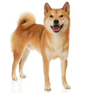
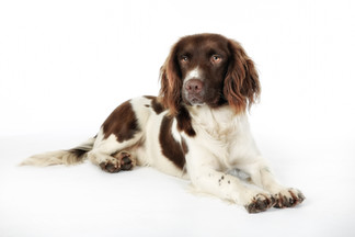
Typically, the condition is common in middle aged pets but variable. In some breeds the female is more predisposed but this is unclear. The dog breeds commonly predisposed to primary closed-angle glaucoma include the Basset Hound, Japanesse Shiba Inu, Flat coated Retriever, Siberian Husky, American Cocker Spaniel, English Cocker Spaniel, Welsh Springer Spaniel, English Springer Spaniel, Border Collie, Great Dane, Dandie Dinmont Terrier, Hungarian Vizla, Leonberger, Welsh Terrier and the Golden Retriever.


What are the clinical signs of Primary Closed Angle Glaucoma?
Companion animals affected by primary closed-angle glaucoma may exhibit symptoms that include the following:-
Acute onset,
Eye redness (Episcleral congestion),
Pain,
Increased tear production,
Squinting,
Corneal oedema,
Corneal cloudiness (opacity),
Mydriasis,
Reduced or absent light responses,
Goniodysgenesis,
Raised intraocular pressure, and
Even vision loss (blindness).

The condition can progress rapidly and is considered an emergency since the increased intraocular pressure can cause damage to the optic nerve, leading to irreversible vision loss if left untreated.
How is Primary Closed Angle Glaucoma Diagnosed?
Diagnosis of primary closed-angle glaucoma in pets typically involves a thorough eye examination (ophthalmic examination) by a veterinarian, including measurements of intraocular pressure using a tonometer (Tonometry), examination of the drainage angle using a gonioscope (gonioscopy), and evaluation of the optic nerve.

Tonometry, the measurement of intraocular pressure (IOP) readily diagnoses glaucoma. There are two types of tonometers that are commonly used by veterinary ophthalmologists, i.e., The Tone-Pen (applanantion tonometer) and TonoVet (rebound tonometer). The normal intraocular pressure ranges between 10mm HG and 15 mm HG. Acute glaucoma cases have intraocular pressures well above 50 mm Hg.
Gonioscopy, examination of the drainage angle, is performed by first applying topical anesthetic to the ocular surface. Then, a goniolens (such as Koeppe or low vacuum Barkan) is filled with fluid before placing on the ocular surface. The opening to the iridocorneal angle is then viewed with the aid of magnification and illumination, for example with a slit-lamp biomicroscope.
Are there conditions to be differentiated from primary closed-angle glaucoma?
The other eye conditions that need to be differentiated from primary closed-angle glaucoma are:-
Secondary glaucoma due to:
Uveitis,
Lens luxation,
Intraocular tumor or
Intraocuar haemorrhage.
Uveitis
Retinal detachment
Treatment
The prognosis for visual recovery after the onset of acute glaucoma is dismal, but treatment options include both medicinal and surgical procedures.
Emergency treatment:
Primary closed angle glaucoma requires emergency treatment to lower intraocular pressure quickly. The options include:
Intravenous administration of hyperosmotic agents (eg 10% mannitol) which reduces intraocular pressure via an osmotic effect on the vitreous and aqueous humours.
Topical application of prostaglandin analogues (eg Latanoprost, travoprost). Side effects include profound miosis and disruption of the blood-aqueous barrier. For these respective reasons, these drugs should not be used in cases of anterior lens luxation and anterior uveitis.
If these emergency measures fail to reduce intraocular pressure to within normal limits then aqueocentesis (under sedation) should be considered.
Medical management
Prostaglandin analogues (eg latanoprost 0.005%, travoprost 0.004%). Topical prostaglandin analogues are used for longer-term treatment of glaucoma. They increase aqueous outflow and are commonly used 1-3 times daily. They are contraindicated in cases of anterior lens luxation.
Carbonic anhydrase inhibitors, eg Dorzolamide 2% and brinzolamide 1%. Used topically, these agents reduce aqueous humor production by inhibiting carbonic anhydrase within the ciliary epithelium. They are usually used 2-3 times daily.
Beta-blockers, eg Timolol 2.5-5%. These drugs also increase aqueous outflow and are usually used 2-3 times daily.
Analgesia (Pain management):
NSAIDs Analgesia
Opioids Analgesia
Surgical therapy
Surgery may be an option for eyes that still have the potential for vision when topical therapy alone is unable to keep intraocular pressure within acceptable ranges. Surgery's main objective is to maintain or restore vision, but if it is successful, it may eventually allow for a reduction in the frequency of needed topical medications. Surgery works by either increasing the amount of aqueous humor that leaves the eye or reducing how much is produced. Even after a successful surgical treatment, there is frequently a continued requirement for relatively frequent topical medications. Various procedures are used that include:-
1. Cycloablation: refers to the destruction of the ciliary processes which are responsible for aqueous humor production. It is performed transclerally or endoscopically from within the eye,
2. Cyclocryoablation refers to the use of nitrous oxide or liquid nitrogen transclerally to reduce IOP or induce phthisis bulbi. This is often associated with prolonged periods of elevated IOP following the procedure and is thus reserved for non-visual eyes.
3. Cyclophotocoagulation refers to the use of laser energy to destroy the ciliary processes. Transcleral cyclophotocoagulation with both Nd:YAG and diode lasers has been reported (Nasisse et al, 1990 and Cook et al, 1997). Complications are fairly frequent and include uveitis, hyphema, cataract formation and retinal detachment. Endoscopic cyclophotocoagulation is now the preferred treatment with encouraging success (Braset al, 2005) .
4. Gonioimplantation refers to the provision of an alternative route of drainage for aqueous humor other than via the iridocorneal angle. Gonioimplants are either unidirectional and valved or bidirectional and non-valved. They are usually positioned episclerally under a dorsal bulbar conjunctival flap. The tubing component of the implant is inserted into the anterior chamber via the sclera taking care to avoid contact with the corneal endothelium, iris and lens .Following placement aqueous humor may exit the anterior chamber via the tubing to form a subconjunctival bleb from where it may be absorbed by the adjacent vasculature. The main complications of gonioimplants are occlusion of the tubing by fibrin and fibrosis around the subconjunctival bleb which may make the utility of the shunt rather short-lived.
5. Eye enucleation and evisceration refers to the enucleation or evisceration with intrascleral prosthesis when there is no hope of restoration of vision despite medical therapy and the eye remains blind and painful.
How is Primary Closed-Angle Glaucoma Prevented?
Although it is unclear what the optimum course of action would be, it has been demonstrated that prophylactic therapy of the other eye can postpone the onset of primary glaucoma (Miller et al., 2000). The majority of ophthamologists recommend applying topical beta-blockers or carbonic anhydrase inhibitors (or both). Topical prostaglandin analogues are also occasionally used, however they may not be the ideal option because they promote shallowing of the anterior chamber and the iridocorneal angle (Tsai et al., 2013).
Prior to breeding, gonioscopy should be used to look for signs of goniodysgenesis in dogs of breeds at risk for primary closed angle glaucoma. A DNA test for Border Collies (OLFML3 mutation, Animal Health Trust) should also always be performed to check for glaucoma. The breeding population should be rid of all canines diagnosed with glaucoma.
References
Pearl R, Gould D, Speiss B (2015) Progression of pectinate ligament dysplasia over time in two populations of Flat-Coated Retrievers. Vet Ophthmol 18, 6-12 PubMed.
Tsai et al (2013) The effect of topical latanoprost on anterior segment anatomic relationships in normal dogs. Vet Ophthmol 16 (5), 370-376 PubMed.
Miller P E et al (2000) The efficacy of topical prophylactic antiglaucoma therapy in primary closed angle glaucoma in dogs: a multicenter clinical trial. JAAHA 36 (5), 431-438 PubMed.
Nasisse M P, Davidson M G, English R V et al (1990) Treatment of glaucoma by use of transcleral neodymium:yttrium aluminium garnet laser cyclophotocoagulation in dogs. JAVMA 197, 350-353 PubMed.
Bras I D, Robbin T E, Wymanet al(2005) Diode endoscopic cyclophotocoagulation in canine and feline glaucoma(abstract). 36th Proceedings of the American College of Veterinary Ophthamologists, p 50.
Cook C S (1997) Surgery for glaucoma. Vet Clin North Am Small Anim Practice 27, 1109-1129.


Comments