


Conn's Syndrome in Cats - When the Adrenal Gland Overshoots

Updated: Aug 16
What is Conn's Syndrome?
Primary hyperaldosteronism, commonly known as Conn’s disease or Conn's syndrome, is an autonomous overproduction of aldosterone from the zona glomerulosa of the adrenal cortex. While once considered exceptionally rare, it is now recognised as the most common adrenocortical disease in domestic cats.

Conn's syndrome is in many ways the mirror image of Addison's disease. Where hypoadrenocorticism starves the body of adrenal hormones, Conn's syndrome floods it with one hormone in particular—aldosterone—secreted independently of the body's normal regulatory signals. It is the most common adrenocortical disease of cats, though it remains widely underdiagnosed because its two hallmark abnormalities, high blood pressure and low blood potassium, are so often attributed to chronic kidney disease instead (Kooistra, 2020).
The condition takes its name from Jerome Conn, the American endocrinologist who first described it in a human patient in 1955. In cats, it is most often caused by a benign or malignant tumour of the adrenal cortex, though in a meaningful minority of cases both adrenal glands are simply overactive without a discrete mass (bilateral hyperplasia) (Kooistra, 2020).
Adrenal Anatomy—Where Aldosterone Is Made
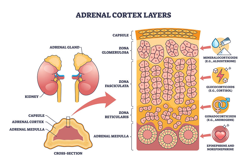
Each cat has two adrenal glands, small and paired, sitting just in front of the corresponding kidney rather than on top of it as in some species. Each gland is really two organs fused into one: an inner medulla that produces adrenaline-type hormones, wrapped in an outer cortex arranged in three concentric layers. Only the outermost of those layers, the zona glomerulosa, is built to manufacture aldosterone. It is this one thin rind of tissue — normally a minor contributor to the gland’s overall bulk — that becomes the source of disease in Conn’s syndrome, whether because a tumour has arisen within it or because the whole layer has become abnormally active (Van Vertloo, 2024).
Adrenal Gland Physiology:
The Feedback Loop Aldosterone Is Meant to Obey

Aldosterone is normally released from the outermost layer of the adrenal cortex, the zona glomerulosa, under the control of the renin–angiotensin–aldosterone system (RAAS). When blood pressure or blood sodium falls, the kidneys release renin, which triggers a cascade culminating in aldosterone release; aldosterone then signals the kidneys to retain sodium (and water) and excrete potassium, restoring blood pressure and volume. Once that goal is met, renin release falls, and aldosterone production settles back down — a self-limiting loop.
In Conn's syndrome, that feedback loop is bypassed entirely. An adrenal tumour, or overactive hyperplastic tissue, secretes aldosterone on its own terms, regardless of what the kidneys or blood pressure are actually signalling. The result is autonomous excess of hormones: blood pressure climbs, and potassium falls, but the gland keeps producing aldosterone anyway because it is no longer listening to the system that is supposed to control it (Kooistra, 2020).
Figure 4. Normal aldosterone release is shut off once blood pressure and sodium are restored. In Conn's syndrome, the adrenal tissue secretes aldosterone independently of this feedback, so the excess never resolves on its own.
Pathophysiology of Feline Conn's Disease
Conn's disease in cats is primarily caused by functional unilateral adrenocortical adenomas, carcinomas, or idiopathic bilateral nodular hyperplasia. The pathognomonic mechanism involves unregulated hypersecretion of aldosterone independent of the renin-angiotensin-aldosterone system (RAAS). Under physiological conditions, aldosterone balances systemic fluids by reclaiming sodium and excreting potassium (Kooistra, 2020)
In a pathological state, excessive aldosterone forces massive renal wasting of potassium into the urine, resulting in profound hypokalaemia (typically < 3.0 mEq/L). Chronic hypokalaemia shifts the resting membrane potential of skeletal muscle cells, inducing a severe, generalised hypokalaemic polymyopathy (Rijnberk et al., 2001).
Why an Excess of One Hormone Causes So Many Signs

Because that regulatory brake is missing, aldosterone keeps acting on the kidney’s distal nephron—the last stretch of tubule before urine is finalised—telling it to keep pulling sodium back into the bloodstream while pushing potassium out into the urine. The two consequences follow directly: sodium and water retention drive blood pressure up, while ongoing potassium loss drains the body’s stores of an ion that muscle and nerve tissue depend on (Kooistra, 2020).
Clinical Signs
The clinical picture of Conn's syndrome follows directly from aldosterone's two jobs—retaining sodium and wasting potassium—taken to excess. Most affected cats are middle-aged to older, without a strong breed or sex predisposition (Kooistra, 2020).
Because chronically high blood pressure tends to creep up gradually, many owners notice nothing unusual until the pressure has already damaged a target organ — classically the eye, where a sudden retinal detachment can cause abrupt, irreversible blindness and is often the very reason the cat is brought in for examination (Ash et al., 2005).

The separate problem of chronic potassium depletion (chronic hypokalaemia) produces its own signature signs or recognisable pattern: general muscle weakness; a hallmark (characteristic) downward curl of the neck (cervical ventroflexion) from weakened neck muscles; and a stiff, flat-footed gait (plantigrade stance). Because these signs overlap so heavily with those of chronic kidney disease — a condition Conn's syndrome can itself also cause or worsen over time — the two are easily mistaken for one another, and finding one does not rule out the other (Kooistra, 2020).
Diagnosis

Any cat presenting with unexplained hypertension, hypokalaemia, or both should prompt consideration of Conn's syndrome, particularly when there is no clear alternative explanation. The single best screening test is the plasma aldosterone-to-renin ratio, which detects aldosterone excess that is disproportionate to what the renin system would justify (Kooistra, 2020; Djajadiningrat-Laanen et al., 2011). Because plasma renin activity assays are not always readily available, measuring aldosterone alongside serum potassium is often used as a practical first step.

Once biochemical suspicion is raised, abdominal ultrasound or CT is used to look for an adrenal mass and to determine whether the disease is unilateral (one gland, usually a tumour) or bilateral (both glands, usually hyperplasia)—a distinction that directly shapes the treatment plan, since only unilateral disease is a good surgical candidate (Kooistra, 2020).
Treatment & Management

Unilateral disease
When imaging confirms a single, resectable adrenal tumour, unilateral adrenalectomy is the treatment of choice and can be curative, though it carries real surgical and anaesthetic risk and requires careful perioperative management of blood pressure and electrolytes (Kooistra, 2020).
Bilateral disease, or cats not suited to surgery
Medical management centres on spironolactone, an aldosterone-receptor antagonist that blocks the hormone's effects at the kidney, often paired with an antihypertensive such as amlodipine and, where needed, oral potassium supplementation to correct hypokalaemia (Kooistra, 2020).
References
Ash, R. A., Harvey, A. M., & Tasker, S. (2005). Primary hyperaldosteronism in the cat: A series of 13 cases. Journal of Feline Medicine and Surgery, 7(3), 173–182. https://doi.org/10.1016/j.jfms.2004.08.003
Djajadiningrat-Laanen, S., Galac, S., & Kooistra, H. (2011). Primary hyperaldosteronism: Expanding the diagnostic net. Journal of Feline Medicine and Surgery, 13(9), 641–650. https://doi.org/10.1016/j.jfms.2011.07.017
Javadi, S., Djajadiningrat-Laanen, S. C., Kooistra, H. S., van Dongen, A. M., Voorhout, G., van Sluijs, F. J., van den Ingh, T. S., Boer, P., & Rijnberk, A. (2005). Primary hyperaldosteronism, a mediator of progressive renal disease in cats. Domestic Animal Endocrinology, 28(1), 85–104. https://doi.org/10.1016/j.domaniend.2004.06.010
Kooistra, H. S. (2020). Primary hyperaldosteronism in cats: An underdiagnosed disorder. Veterinary Clinics of North America: Small Animal Practice, 50(5), 1053–1063. https://doi.org/10.1016/j.cvsm.2020.05.007
MSD Veterinary Manual. (2025). Feline primary hyperaldosteronism. https://www.msdvetmanual.com/endocrine-system/the-adrenal-glands/feline-primary-hyperaldosteronism


Comments