The Growth That Won't Go Away: Keloid Scars in Dogs and Cats
- Dr Andrew Matole, BVetMed, MSc
- Dec 31, 2023
- 5 min read
What are keloid scars in dogs?

A keloid scar, also called a hypertrophic scar, is a thick, raised overgrowth of collagen tissue. They are often shiny and firm to the touch and may have a flesh-colored or dark brown pigment. Keloid scars can look like large, hairless areas around the wound site. They can also look like nodules or masses under the skin and may be indistinguishable from other types of growths in dogs.

Keloids in dogs and cats, as in humans, are abnormal growths of fibrous tissue that occur at the site of a wound or injury. These growths are characterized by their excessiveness and the fact that they extend beyond the boundaries of the original wound. While keloids in pets do occur, they are relatively rare compared to their occurrence in humans.


A keloid scar on a dog's foot after surgery A hypertrophic scar on a child's elbow
Keloids in dogs and cats are relatively uncommon, but they are a specific type of abnormal skin growth that can occur in some breeds. A keloid is a raised, thickened area of scar tissue that extends beyond the boundaries of the original wound or injury. They can be quite distinct from typical scar tissue and can cause cosmetic and health concerns for pets.
In some cases, keloids can be irritating or even painful causing discomfort or itching for the pet. If the dog or cat scratches at the scar, it could result in a secondary infection. Keloid scars can also be potentially malignant (cancerous) tumours. Keloids can occur anywhere on a pet's body but are most commonly seen in areas with minimal subcutaneous fat, such as the chest, ears, elbows, knees and shoulders.
What Causes Keloids?
The exact cause of keloids in animals, as well as in humans, is not fully understood. However, genetic factors, breed predisposition, an individual's propensity to develop excessive scar tissue and the type of wound or incision made during surgery may all play a role and contribute to their formation. Keloids can develop after surgical procedures, trauma, or even spontaneously in some cases.
How do Keloids Develop?
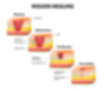
When a dog's skin sustains a lesion or bite, the body initiates a natural healing process. As part of this process, a protein called collagen is released to help close the wound and regenerate damaged tissue. This creates a scar. Scar tissue forms in almost all wounds except for the most minor cuts and scratches. While most types of scars are harmless, keloid scars are different.

Keloids are a result of an overproduction of collagen, which is a protein involved in wound healing. When there is an excessive production of collagen in response to an injury or surgery, it leads to the formation of a keloid.

Keloids can vary in size, shape, and colour, but they typically appear as raised, thickened, shiny and irregularly shaped lumps on the skin. They may appear reddish, pink, purplish, or darker than the surrounding skin and they often continue to grow beyond the original wound size.

Keloidal scarring can indicate tumors, known as keloidal fibromas or fibrosarcomas, that require surgical removal and monitoring. Keloidal fibromas are uncommon in dogs, and other animal species are not affected. The relative rarity of keloidal lesions in animals contrasts with the common occurrence of keloidal lesions (keloids and hyperplastic scars) in humans.

The presence of macrophages in most keloidal tumors suggests that in dogs, as in humans, keloidal fibromas and fibrosarcomas develop secondary to tissue injury and may represent a reactive inflammatory lesion rather than a true neoplasm.
How are Keloid scars different from other types of scars in dogs?
Keloid scars usually expand beyond the original wound site. Other scar types may change color or thickness but do not grow or extend beyond the original wound. Also, whereas most scars fade with time, keloid scars often reappear after removal.
Keloid scars are also different histologically, or in their microscopic structure. They contain thickened bundles of hyalinized collagen fibers that differentiate them from other collagen-based nodules and lesions. Hyalinization occurs when normal tissue deteriorates into a homogeneous, clear material. Also, unlike other scars, keloids have more inflammatory cells and often more vascularized.

Hyalinized collagen fibers are a distinctive feature of canine keloidal tumors and of keloids and hypertrophic scars in humans. Canine keloidal lesions also differ from hypertrophic scars in that they contain more hyalinized collagen fibers and are not linear lesions located at the site of previous surgery. Thus, keloidal lesions in dogs histologically resemble but are distinct from keloids and hypertrophic scars of humans.
What are the Risk Factors for Keloids in dogs and cats?
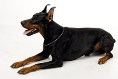
Keloids are more commonly observed in certain breeds that are predisposed to develop excessive scar tissue. Some dogs and cats may be more prone to keloid formation due to their breed, age, or genetic background. Certain breeds, such as Boxers, Doberman Pinschers, Bull Terriers and Siamese cats, are more predisposed to keloids.


Epidemiologically, male dogs are more often affected by keloidal tumors than females, whereas keloidal lesions in humans predominantly affect females. There is no age predominance in dogs with keloidal tumors, whereas keloidal lesions predominantly develop in young adults in humans. These epidemiologic differences further suggest that keloidal tumors in dogs are not the exact counterpart of keloidal lesions in humans.
Canine keloidal lesions have distinctive histologic features with the possibility of transformation from canine keloidal fibromas into malignant fibrosarcomas.
What is the Treatment for Keloids in dogs and cats?
Managing keloids in pets can be challenging and bothersome for pets as they may itch or cause discomfort. Treatment options include surgical removal, laser therapy, steroid injections, silicone gel sheeting, or a combination of these approaches. However, it's important to note that keloids can be stubborn and may recur even after treatment. Keloidal fibromas and fibrosarcomas typically develop slowly, therefore, early intervention can improve the likelihood of a successful treatment.

Scar treatment for keloids varies depending on whether it is benign or malignant. Once the scar has been examined by a veterinarian and a biopsy of the area taken to determine if it is a keloidal fibrosarcoma or not, then portions of the tissue can be removed if it is a benign fibroma as complete excision is generally not recommended for benign scar tissues. Complete excision of benign scar tissues increases the chance of the scar reoccurring.
Corticosteroids, such as methylprednisolone acetate, may be used to slow the scar's growth. These can be injected into the scar tissue. Topical skin treatments may also be prescribed as well to alleviate pain, inflammation, and irritation. While these interventions do not eliminate the keloid, they can reduce discomfort and the risk of infection. The dog may need multiple treatments, as keloid scars tend to regenerate.
If the scar tissue is malignant, a complete surgical removal is necessary to completely stop the cancer from spreading.
What is the Prevention of Keloids in dogs and cats?
Since the exact cause of keloids is not completely understood, it can be difficult to prevent their formation. However, minimizing trauma to the skin during surgical procedures and closely monitoring wounds for signs of excessive scarring can be helpful. To reduce the risk of keloid formation in pets, it's essential to minimize unnecessary surgery and ensure proper wound care. This may include using techniques to minimize tissue trauma during surgery and post-operative wound management.
References
Cullen, J. M. (2008). Tumors of the Skin and Soft Tissues. In Withrow and MacEwen's Small Animal Clinical Oncology (4th ed.). Saunders.
Goldschmidt, M. H., Goldschmidt, K. H., & Hendrick, M. J. (2002). Proliferative cutaneous lesions of the dog and cat. In Skin Tumors of the Dog and Cat (pp. 79-134). Pergamon.
Mikaelian I, Gross TL. Keloidal Fibromas and Fibrosarcomas in the Dog. Veterinary Pathology. 2002;39(1):149-153. doi:10.1354/vp.39-1-149
Muller & Kirk's Small Animal Dermatology by William H. Miller Jr. and Craig E. Griffin.
Veterinary Surgery: Small Animal by Karen M. Tobias and Spencer A. Johnston.
Journal of Small Animal Practice
Journal of Veterinary Dermatology