Tricuspid valve dysplasia (TVD)
- Dr Andrew Matole, BVetMed, MSc

- May 21, 2023
- 4 min read

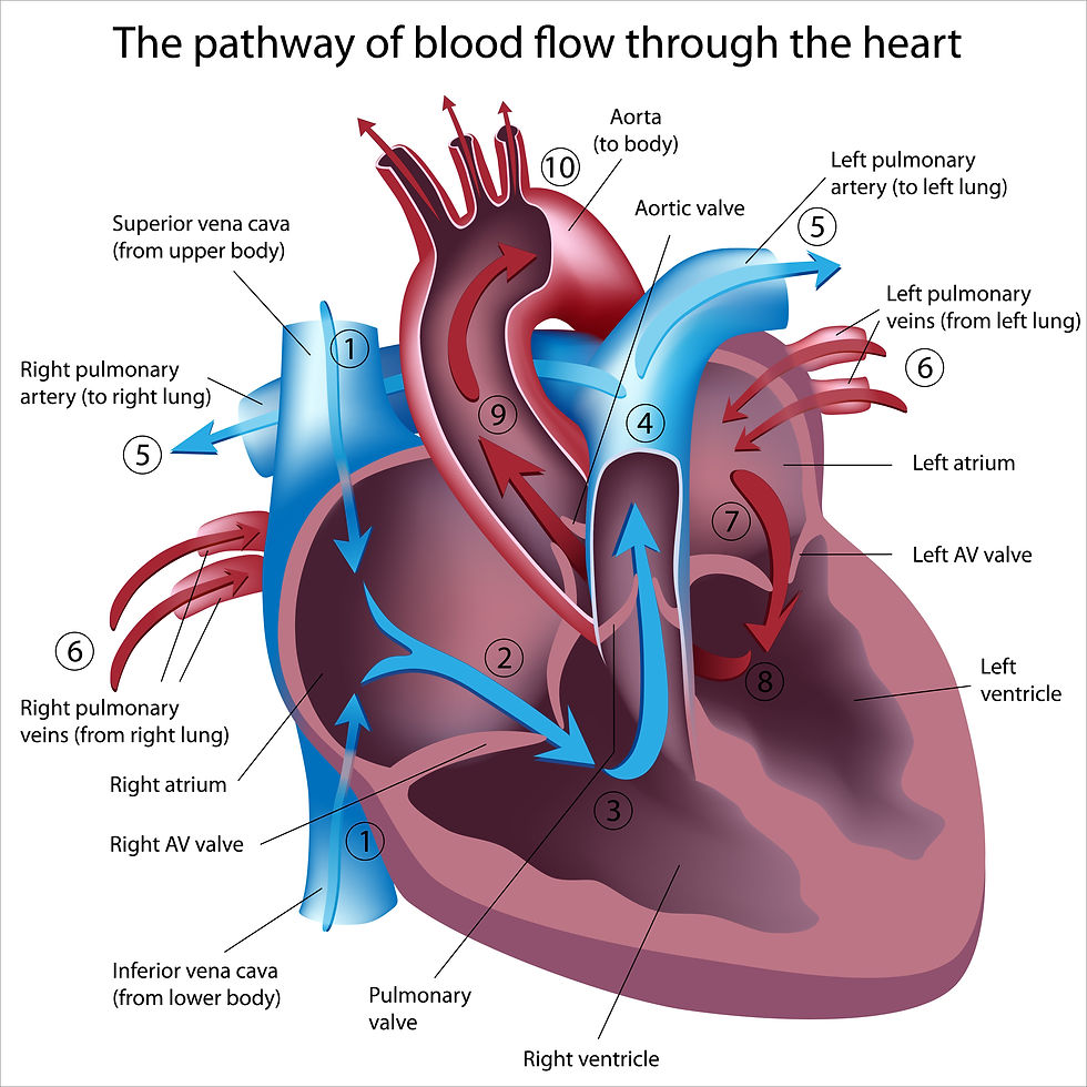
Tricuspid valve dysplasia, which affects both humans and canines, is a congenital cardiac disorder involving the tricuspid valve and its accompanying structures (such as papillary muscles and chordae tendineae). It involves abnormal growth of the tricuspid valve, which is situated between the right atrium and the right ventricle of the heart. Three leaflets make up the tricuspid valve, which opens and closes to control blood flow between these chambers. According to one study, it makes up 2% of all congenital diseases in dogs, making it a rare congenital cardiac issue. Large breed dogs, and the Labrador Retriever in particular, are the breeds where TVD is most frequently found. Other breeds commonly involved are Boxers, German Shepherd Dogs, Dougue de Bordeaux and Great Danes. Cats can infrequently develop TVD. Tricuspid regurgitation, right heart hypertrophy, and in severe cases, right heart failure are also symptoms of TVD.

In both dogs and humans, tricuspid valve dysplasia refers to the malformation of the valve leaflets, leading to improper functioning. The severity of the dysplasia can vary, ranging from mild cases where the valve is slightly malformed to severe cases where the valve is completely deformed or even absent.
When a cardiac murmur is picked in young puppies or kittens, tricuspid valve dysplasia (TVD) is predisposed, albeit the animal frequently shows no symptoms. However, many animals continue to be asymptomatic for years. Clinical symptoms do rise as the animal ages.
How does Tricuspid Valve Dysplasia Clinically Present?

Clinical symptoms frequently manifest as an asymptomatic murmur, and animals might go for years without showing any symptoms. The presence of ascites, jugular pulsation, pleural effusion, labored breathing caused by the compromise of the thoracic space caused by ascites, weakness, syncopal episodes (poor cardiac output secondary to, for example, dysrhythmia and/or congestive heart failure), and poor appetite are all signs of right heart failure that typically predominate in severely affected animals.
How is Tricuspid Valve Dysplasia Diagnosed?
The diagnosis of tricuspid valve dysplasia is done through a good client history, a thorough physical examination to pick up heart murmurs on routine exam and presenting clinical signs of congestive heart failure including weakness, lethargy, abdominal distension and subsequent unexplained weight gain. Other clinical sign to be noted is poor appetite and syncopal episodes.
Diagnostic investigations should include chest radiographs (x-rays) and cardiac ultrasound. Electrocardiography (ECG) can also be done including abdominal tap and fluid analysis of ascites plus central venous pressure. Abdominal fluid tap analysis in cases of TVD show a modified transudate consistent with heart failure.

Chest radiographs: right atrial and ventricular enlargement is mostly present, often times dramatic and severe, enlarged caudal vena cava . Right atrial enlargement. There is also distension of the caudal vena cava in right-sided congestive heart failure (CHF) including pleural effusion. hepatomegaly and ascites.

Electrocardiogram (ECG): tall P waves (p - pulmonale), mean electrical axis shift to the right and criteria for right ventricular enlargement (deep S waves). Splintered intraventricular conduction disturbances and particularly atrial fibrillation are commonly found in TVD.
Elevated central venous pressure (CVP) means the presence of high systemic venous pressures and right heart failure. In the presence of ascites, characteristic ultrasound findings and clinical signs, CVP measurements are often not required to formulate a diagnosis.
How is the post-mortem of a Tricuspid Valve Dysplasia?
At post-mortem, the tricuspid valve leaflet displays varying amounts of thickening, with the chordae tendineae shortened, occasionally to the point of being non-existent. In these cases the valve leaflet attaches directly to the papillary muscles . The papillary muscles are often malpositioned and malformed. Occasionally, only a single papillary muscle is found. The valve leaflets may be partially adhered to the ventricular or septal wall. The right atrium and ventricle display eccentric hypertrophy.
How is Tricuspid Valve Dysplasia Managed?
The management of tricuspid valve dysplasia in both dogs and humans depends on the severity of the condition and the presence of associated heart defects. Treatment may involve medication to manage symptoms and improve heart function, surgical repair or replacement of the valve, or other interventions aimed at alleviating the effects of the disease. However, the basic goals of the treatment for congestive heart failure should be followed:
Reduction of preload - using venodilators and diuretics.
Reduction of afterload - using arteriodilators.
Positive inotropic support.
Treatment of complications, eg arrhythmias, cardiac cachexia
References
Ohad DG, Avrahami A, Waner T, David L. The occurrence and suspected mode of inheritance of congenital subaortic stenosis and tricuspid valve dysplasia in Dogue de Bordeaux dogs. Vet J. 2013 Aug;197(2):351-7. doi: 10.1016/j.tvjl.2013.01.012. Epub 2013 Feb 20. PMID: 23434219.
Hoffmann G, Amberger CN, Seiler G, Lombard CW. Trikuspidaldysplasie bei fünfzehn Hunden [Tricuspid valve dysplasia in fifteen dogs]. Schweiz Arch Tierheilkd. 2000 May;142(5):268-77. German. PMID: 10850163.
Liu SK, Tilley LP. Dysplasia of the tricuspid valve in the dog and cat. J Am Vet Med Assoc. 1976 Sep 15;169(6):623-30. PMID: 134984.
Atkins C, Bonagura J, Ettinger S, Fox P, Gordon S, Haggstrom J, Hamlin R, Keene B, Luis-Fuentes V, Stepien R. Guidelines for the diagnosis and treatment of canine chronic valvular heart disease. J Vet Intern Med. 2009 Nov-Dec;23(6):1142-50. doi: 10.1111/j.1939-1676.2009.0392.x. Epub 2009 Sep 22. PMID: 19780929.


Comments