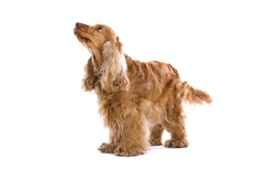
Understanding Dandruff in Pets: Causes, Types, and Clinical Relevance
- Dr Andrew Matole, BVetMed, MSc

- May 31, 2025
- 11 min read
Updated: Jun 9, 2025
What is Dandruff in Pets?

Dandruff in pets refers to the noticeable shedding of dead skin cells, often seen as white or greyish flakes on the fur or bedding. This dermatologic issue is prevalent in both dogs and cats, though its severity and appearance can vary depending on the animal's breed, health status, environment, and grooming habits (Scott, Miller, & Griffin, 2001).
Although often perceived as a superficial or cosmetic issue, dandruff can sometimes be an early indicator of underlying systemic or dermatologic disease. Environmental factors such as low humidity or sudden dietary changes may cause transient flaking. However, persistent or severe dandruff may reflect deeper health conditions that require veterinary diagnosis and management (Miller, Griffin, & Campbell, 2013).
Skin disorders are among the most common reasons for veterinary consultations. A study published in the Journal of Veterinary Dermatology reported that approximately 26% of dogs and cats present with dermatologic issues, many of which are associated with scaling and seborrhea (Bond et al., 2020).
What Is Seborrhea in Pets?

Clinically, dandruff is a symptom of seborrhea, a disorder characterised by abnormal keratinisation and/or sebum production. It results in the accumulation of visible skin flakes (Scott, Miller, & Griffin, 2001; Moriello, 2018). Keratinisation is the process where new skin cells form, move to the surface, harden, and eventually shed as dead skin. It helps keep the skin healthy and protected, but when disrupted, it can lead to dandruff or other skin issues (Miller, Griffin, & Campbell, 2013). The flakes or dandruff can further be classified based on the type of scaling and oiliness of the skin:

Seborrhea Sicca (Dry Seborrhea): Presents with dry, powdery, white flakes and a dull coat appearance. It resembles human dandruff and often results in mild itching (Scott, Miller, & Griffin, 2001).
Seborrhea Oleosa (Oily Seborrhea): Characterised by greasy, yellowish scales, excessive oil production, and a distinctive rancid odour. The skin and coat may appear sticky and matted (Miller, Griffin, & Campbell, 2013).

Mixed Seborrhea: Many pets display features of both sicca and oleosa, particularly when secondary infections or systemic conditions are involved.
Table I: Summary Table of differences between dandruff and seborrhoea
Feature | Dandruff | Seborrhoea |
Type | Symptom | Medical condition |
Appearance | White/grey dry flakes | Dry flakes, greasy skin, or both |
Cause | Mild irritation, dry skin | Keratinisation/sebum disorder |
Severity | Usually mild | Often chronic and progressive |
Diagnosis | Visual observation | Clinical diagnosis |
Treatment Focus | Hydration, grooming, basic care | Medical therapy targeting the root cause |
Primary vs. Secondary Seborrhoea
Seborrhea can be either primary or secondary, with secondary being far more common.
Primary Seborrhea is a hereditary keratinisation disorder that typically affects specific breeds such as Cocker Spaniels, West Highland White Terriers, Basset Hounds, and Dachshunds. This form is congenital and often lifelong (Miller, Griffin, & Campbell, 2013).
Secondary Seborrhea results from other underlying health problems such as endocrine disorders, parasitic infestations, nutritional imbalances, allergies, or infections. In such cases, resolving the root cause is essential for controlling the dandruff (Moriello, 2018).


It is essential to understand that seborrhea is not a standalone disease but rather a clinical manifestation of systemic, environmental, or dermatologic imbalances.
Causes of Dandruff in Pets

Identifying the underlying cause of dandruff in pets is critical for determining the most effective treatment and ensuring long-term skin health. Dandruff, often a symptom rather than a standalone condition, can result from several contributing factors, ranging from environmental conditions to systemic disease.
Allergies

Both environmental and food allergies can lead to chronic skin inflammation, which disrupts normal skin function and leads to flaking or dandruff. Environmental allergens such as pollen or dust mites can cause atopic dermatitis, while food allergies may trigger localised or generalised skin issues. Approximately 10% of dogs experience food allergies that manifest through dermatologic signs (Marsella, Olivry, & Carlotti, 2011; Hillier & Griffin, 2001).
Skin Disorders

Conditions such as seborrhea, dermatitis, and fungal infections are primary dermatological causes of dandruff. Accurate diagnosis is essential, as treatment varies widely depending on the condition. Seborrheic dermatitis, for example, involves abnormalities in keratinisation and/or sebum production (Scott, Miller, & Griffin, 2001), while fungal infections like Malassezia dermatitis require antifungal therapy (Bond et al., 2020).
Nutritional Deficiencies

Diet plays a significant role in maintaining skin health. Inadequate intake of essential fatty acids (particularly omega-3 and omega-6), zinc, and vitamin A can lead to dry, flaky skin and impaired skin barrier function (Watson, 1998). Supplementation with omega-3 fatty acids has been shown to reduce skin inflammation by up to 30% in affected animals (Logas & Kunkle, 1994).
Environmental Conditions

Low humidity, especially in winter or in homes with central heating, dries out the skin and exacerbates dandruff. Seasonal changes and rapid temperature shifts can impair the skin's ability to retain moisture (Miller, Griffin, & Campbell, 2013; Moriello, 2018)
Parasitic Infections

External parasites such as Cheyletiella mites (nicknamed “walking dandruff”), fleas, lice, and demodex mites can irritate the skin and cause or worsen scaling. These parasites lead to hypersensitivity reactions, pruritus, and secondary infections (Mueller et al., 2020; Scott, Miller, & Griffin, 2001).
6. Infections

Both bacterial (pyoderma) and fungal infections can significantly affect skin integrity. Bacterial infections cause inflammation, scaling, and crusting, while Malassezia yeast and dermatophytes (e.g., ringworm) result in greasy, flaky skin, especially in moist areas like skin folds (Bond et al., 2020; Miller, Griffin, & Campbell, 2013).
7. Endocrine (Hormonal) Disorders

Systemic diseases such as hypothyroidism and Cushing's disease (hyperadrenocorticism) often manifest through skin and coat changes. Hypothyroidism typically causes dry, brittle hair and scaling, while Cushing's may lead to thinning skin and secondary infections that contribute to dandruff (Beale, 2001; Behrend, Kooistra, & Nelson, 2013).
8. Poor Grooming or Mobility Issues

Owners' Infrequent or improper grooming and over-bathing with harsh shampoos can also compromise the skin barrier (Hensel, 2010).

In animals that are obese, arthritic, or in pain, grooming behaviour may be reduced. This is particularly common in older cats, leading to the accumulation of dead skin and debris on the coat (Buffington, 2002).
9. Systemic Illness

Chronic internal diseases such as liver disease, neoplasia, or autoimmune disorders may present dermatologic signs, including seborrhea or scaling. These cases require a comprehensive diagnostic evaluation (Miller, Griffin, & Campbell, 2013).
Table II: Summary Table for Common Causes of Pet Dandruff
Cause | Examples |
Allergies | Atopy, food allergies, flea allergy dermatitis |
Skin Disorders | Seborrhea, dermatitis, Malassezia dermatitis |
Nutritional Deficiencies | Low omega-3, zinc, and vitamin A |
Environmental Factors | Dry air, low humidity, and indoor heating |
Parasites | Cheyletiella mites, fleas, lice, and demodex |
Infections | Bacterial pyoderma, fungal infections (ringworm, Malassezia) |
Endocrine Disorders | Hypothyroidism, Cushing’s disease |
Grooming Issues | Obesity, arthritis, over-/under-bathing, poor grooming routines |
Systemic Illness | Liver disease, cancers, and autoimmune skin disorders |
Clinical Importance and Next Steps
While flakes on a pet’s coat or bedding may initially seem minor, they often reflect more than just dry skin. Dandruff can be a marker of poor nutrition, endocrine dysfunction, external parasites, or chronic skin inflammation.
Veterinarians are crucial in distinguishing between simple environmental causes and more serious underlying diseases. Diagnostic tools such as skin cytology, blood panels, and allergy testing are often necessary to identify the root cause and guide treatment.
This guide aims to help pet owners recognise signs of dandruff early, understand its clinical context, and pursue appropriate care strategies for promoting healthier skin and overall well-being in their pets.
Diagnosing Dandruff in Pets
Noticing flakes on your pet’s coat should prompt a veterinary consultation, as dandruff is often a clinical sign of an underlying problem, not a disease in itself. Accurate diagnosis is key to formulating an effective treatment plan, especially since most cases of dandruff are linked to secondary seborrhea, which arises from other health conditions rather than occurring in isolation (Scott, Miller, & Griffin, 2001).
Initial Veterinary Assessment
Diagnosis typically begins with:
Comprehensive History

The veterinarian will gather detailed information on the pet’s:
Diet and supplements
Grooming practices and bathing frequency
Parasite prevention measures
Recent environmental changes
Onset, duration, and progression of dandruff
Signs of itching or discomfort
This contextual background helps narrow down potential causes and guide the diagnostic process (Miller, Griffin, & Campbell, 2013).
Physical Examination

A veterinary doctor conducting a physical examination A thorough physical evaluation focuses on:
Location and pattern of scaling
Type of flakes (dry vs. oily)
Presence of odour, inflammation, or crusts
Hair coat quality and signs of hair loss
Evidence of parasitic infestations or infections
Diagnostic Tests
To confirm the diagnosis and identify the root cause, the following diagnostic tools may be employed:
1. Skin Scrapings
Used to detect external parasites such as Demodex or Sarcoptes mites.
2. Tape Impressions and Skin Cytology
Help identify surface bacteria, Malassezia yeast, or parasitic elements like Cheyletiella mites (Moriello, 2018).
3. Fungal Cultures
Essential for detecting dermatophytosis (ringworm), especially in multi-pet households or zoonotic risk cases.
4. Bacterial Culture and Sensitivity Testing

Conducted when pyoderma is suspected, allowing for targeted antibiotic therapy.
Advanced Investigations (As Needed)
If the basic diagnostics are inconclusive or if a chronic or systemic cause is suspected, further testing may include:
Allergy Testing
Through intradermal skin testing or serum IgE testing to identify atopic dermatitis.
Food Elimination Trials
To determine food-related hypersensitivities by feeding a limited-ingredient or novel protein diet for 8–12 weeks (Hillier & Griffin, 2001).
Blood Tests

Complete blood count (CBC) and biochemistry panel
Thyroid function tests (to detect hypothyroidism)
Adrenal function tests (e.g., ACTH stimulation test) to assess for Cushing’s disease
Skin Biopsy

Recommended in chronic or complex cases where autoimmune skin disease, neoplasia, or unusual dermatoses are suspected (Scott, Miller, & Griffin, 2001).
Table III: Summary of Dandruff / Seborrhoea
Purpose | |
History & Physical Exam | Identify potential external or lifestyle-related factors |
Skin Scrapings | Check for mites (e.g., Demodex, Sarcoptes) |
Tape Cytology | Detect bacteria, yeast, or Cheyletiella |
Fungal Culture | Confirm dermatophytosis (ringworm) |
Blood Tests | Rule out hormonal and systemic diseases |
Food Trials | Investigate dietary allergies or intolerances |
Skin Biopsy | Diagnose autoimmune or complex skin conditions |
Treatment and Management of Dandruff in Pets

Successfully managing dandruff in dogs and cats requires a comprehensive, two-pronged approach:
Targeting the underlying cause, especially when dandruff is secondary to a deeper pathology
Providing symptomatic relief reduces flaking, supports skin barrier function, and enhances comfort.
Addressing the Root Cause
In most cases, dandruff stems from secondary seborrhea, which is triggered by an underlying condition. Accurate diagnosis and targeted treatment are essential. Common underlying conditions and their interventions include:
Parasitic Infestations


Mites such as Cheyletiella, Demodex, and fleas can lead to significant skin irritation and scaling. Effective control involves topical or systemic antiparasitic medications (Mueller et al., 2020).
Infections
Bacterial pyoderma may require antibiotics, while fungal infections like Malassezia dermatitis or dermatophytosis (ringworm) respond to antifungals such as ketoconazole or miconazole (Bond et al., 2020).
Allergies


Pets with food allergies or environmental sensitivities may benefit from antihistamines, corticosteroids, or immunotherapy. Sometimes, an elimination diet helps identify and remove dietary triggers (Marsella, Olivry, & Carlotti, 2011).
Hormonal Imbalances


Endocrine disorders like hypothyroidism and Cushing's disease often present with dermatological signs, including scaling. Treatment may involve levothyroxine for thyroid deficiency or trilostane for hyperadrenocorticism (Beale, 2001; Behrend, Kooistra, & Nelson, 2013).
Symptomatic Relief Through Topical Therapy
Topical treatments help manage visible flakes, soothe inflammation, and restore the integrity of the skin barrier. Selection of products should be tailored to the type (dry vs. oily) and severity of seborrhea (Miller, Griffin, & Campbell, 2013).
Standard Classes of Topical Ingredients:
Keratolytic and Keratoplastic Agents

Agents such as salicylic acid, sulfur, and coal tar promote exfoliation and normalise skin cell turnover. Note: Coal tar should be avoided in cats due to potential toxicity.
Degreasing Agents

Benzoyl peroxide and selenium sulfide are effective in managing oily seborrhea. However, they may be too drying if used too frequently or on already compromised skin.
Antimicrobials


Topical antimicrobials like chlorhexidine, ketoconazole, and miconazole reduce microbial overgrowth, especially in cases involving secondary bacterial or yeast infections (Bond et al., 2020).
Moisturisers and Emollients


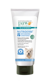


Ingredients such as colloidal oatmeal, ceramides, and essential fatty acids help hydrate the skin, reduce inflammation, and support epidermal repair (Hensel, 2010).
The frequency and type of topical application should be guided by veterinary assessment, considering whether the seborrhea is predominantly dry, oily, or mixed (Miller et al., 2013).
Dietary Supplementation
Supplementing the pet’s diet with omega-3 and omega-6 fatty acids, commonly found in fish oil or marine-based sources, has proven benefits. These fatty acids reduce inflammation, improve skin hydration, and enhance coat quality, especially in pets with allergies or chronic dry skin (Logas & Kunkle, 1994; Watson, 1998).


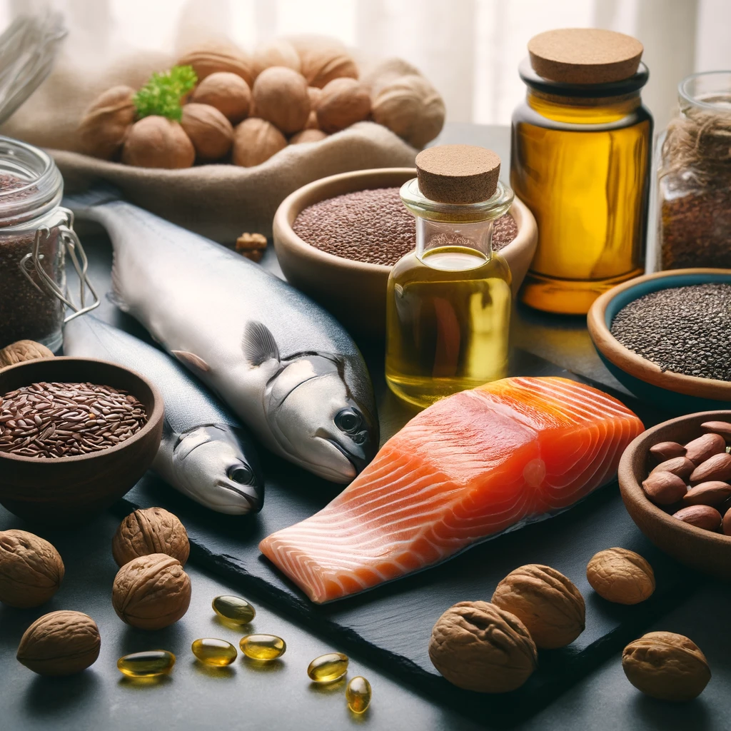


Table IV: Summary of Management Strategies for Dandruff and Seborrhoea
Management Area | Examples | References |
Underlying Cause | Parasite control, antimicrobial therapy, endocrine or allergy treatment | Beale, 2001; Bond et al., 2020; Mueller et al., 2020 |
Medicated Shampoos | Salicylic acid, sulfur, selenium sulfide, benzoyl peroxide | Miller et al., 2013 |
Antimicrobial Agents | Chlorhexidine, miconazole, ketoconazole | Bond et al., 2020 |
Emollients and Moisturizers | Oatmeal, ceramides, essential fatty acids | Hensel, 2010 |
Nutritional Support | Omega-3 and omega-6 supplementation | Logas & Kunkle, 1994; Watson, 1998 |

Preventing Dandruff in Pets
Although primary seborrhea—a genetically inherited disorder of keratinisation—cannot be prevented, secondary seborrhea, which arises from underlying medical or environmental factors, can be minimised through proactive care strategies (Miller, Griffin, & Campbell, 2013).
Key Preventive Measures:
Nutritionally Balanced Diet
Feeding your pet a high-quality diet enriched with essential nutrients, particularly omega-3 and omega-6 fatty acids, supports optimal skin function and reduces the risk of flaking and inflammation (Watson, 1998; Logas & Kunkle, 1994).
Consistent Grooming Practices
Regular brushing helps remove dead skin and loose hair, while stimulating oil glands to promote a healthy, well-lubricated coat. Grooming should be tailored to your pet’s coat type and may include the use of hypoallergenic or medicated shampoos when indicated (Hensel, 2010).
Year-Round Parasite Prevention
Maintaining routine flea and tick prevention reduces the risk of irritation and hypersensitivity reactions that can lead to skin scaling or infections (Mueller et al., 2020).
Prompt Allergy Management
Early identification and treatment of food or environmental allergies help prevent chronic inflammation, a key driver of secondary seborrhea (Marsella, Olivry, & Carlotti, 2011).
Environmental Control
Using a humidifier in dry or air-conditioned environments can help maintain skin hydration and prevent flaking. Ensuring your pet lives in a clean, allergen-controlled space can reduce skin flare-ups (Moriello, 2018).
Routine Veterinary Examinations
Annual or biannual check-ups are crucial for both preventing and managing dandruff. Regular check-ups allow for early detection of endocrine (hormonal) or dermatologic diseases, improving prognosis and reducing the risk of chronic skin conditions developing unnoticed (Beale, 2001).
When to See the Vet
Seek veterinary attention if your pet exhibits any of the following:
Persistent or worsening dandruff
Excessive scratching, licking, or biting
Hair loss, redness, or skin odour
Signs of infection (e.g., pustules, greasy patches, scabbing)
Noticeable discomfort or behaviour changes
While dandruff may appear harmless, not always serious, it can be a symptom of a more serious underlying condition, the tip of an iceberg indicating deeper systemic issues requiring medical evaluation and treatment (Moriello, 2018).
Conclusion
Dandruff in pets—whether dry, oily, or mixed—is rarely just a cosmetic issue. It often reflects deeper disturbances in skin barrier function or internal health, such as infections, allergies, endocrine disorders, or nutritional imbalances. Successful management hinges on accurate diagnosis, targeted treatment, and long-term preventive care. Pet owners play a vital role in monitoring their animal's skin health and should consult a veterinarian at the first sign of persistent flaking or coat changes.
The journey to healthier skin for your pet begins with knowledge and proactive care. When your pet's skin is healthy, they are happier and more active.

Need Help with Your Pet’s Skin?
Book a consultation at The Andys Veterinary Clinics and let our experienced team help your pet look and feel their best—fluff without the flakes!
References
Beale, K. M. (2001). Dermatologic manifestations of endocrine disorders. Veterinary Clinics: Small Animal Practice, 31(5), 1035–1056.
Bond, R., Guillot, J., & Kano, R. (2020). Evidence-based veterinary dermatology: Best practice for Malassezia dermatitis. Veterinary Dermatology, 31(1), 28–34.
Behrend, E. N., Kooistra, H. S., & Nelson, R. (2013). Diagnosis of spontaneous canine hyperadrenocorticism: 2012 ACVIM consensus statement (small animal). Journal of Veterinary Internal Medicine, 27(6), 1292–1304.
Buffington, C. A. T. (2002). External and internal influences on disease expression in cats with feline lower urinary tract disease. Journal of the American Veterinary Medical Association, 220(4), 594–596.
Hensel, P. (2010). Nutrition and skin diseases in veterinary medicine. Clinics in Dermatology, 28(6), 686–693.
Hillier, A., & Griffin, C. E. (2001). The ACVD task force on canine atopic dermatitis (I): incidence and prevalence. Veterinary Immunology and Immunopathology, 81(3-4), 147–151.
Logas, D., & Kunkle, G. (1994). Evaluation of diets and fatty acid supplements in dogs with atopic dermatitis. Veterinary Dermatology, 5(3), 99–104.
Marsella, R., Olivry, T., & Carlotti, D. N. (2011). Current evidence of skin barrier dysfunction in canine atopic dermatitis. Veterinary Dermatology, 22(3), 239–248.
Miller, W. H., Griffin, C. E., & Campbell, K. L. (2013). Muller & Kirk's Small Animal Dermatology (7th ed.). Saunders.
Mueller, R. S., Bensignor, E., & Ferrer, L. (2020). Diagnosis and treatment of ectoparasites in dogs and cats. Compendium: Continuing Education for Veterinarians, 42(1), 16–27.
Scott, D. W., Miller, W. H., & Griffin, C. E. (2001). Muller and Kirk’s Small Animal Dermatology (6th ed.). W.B. Saunders Company.
Watson, T. D. G. (1998). Diet and skin disease in dogs and cats. The Journal of Nutrition, 128(12 Suppl), 2783S–2789S.


Comments